Eye & Ear Anatomy Prints
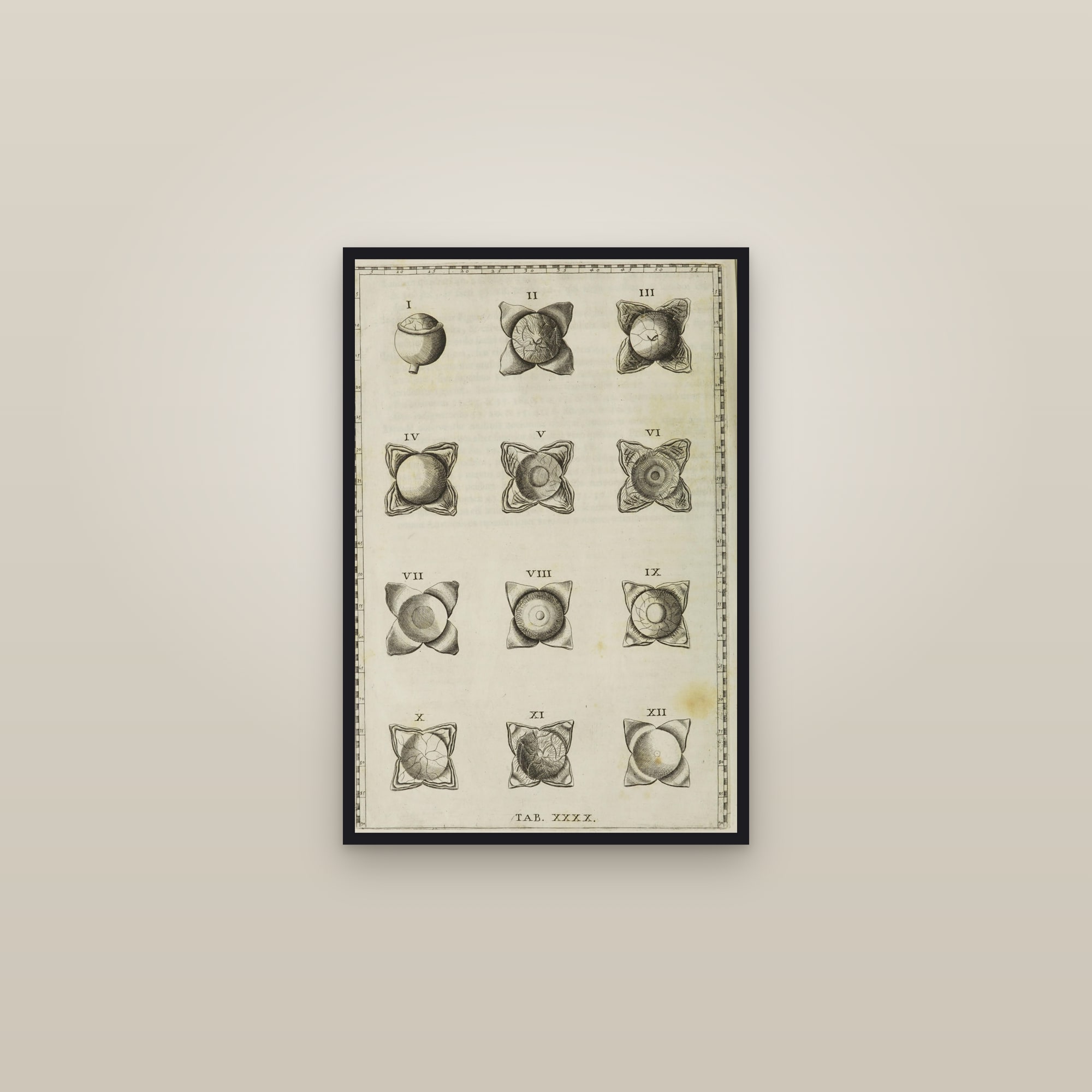
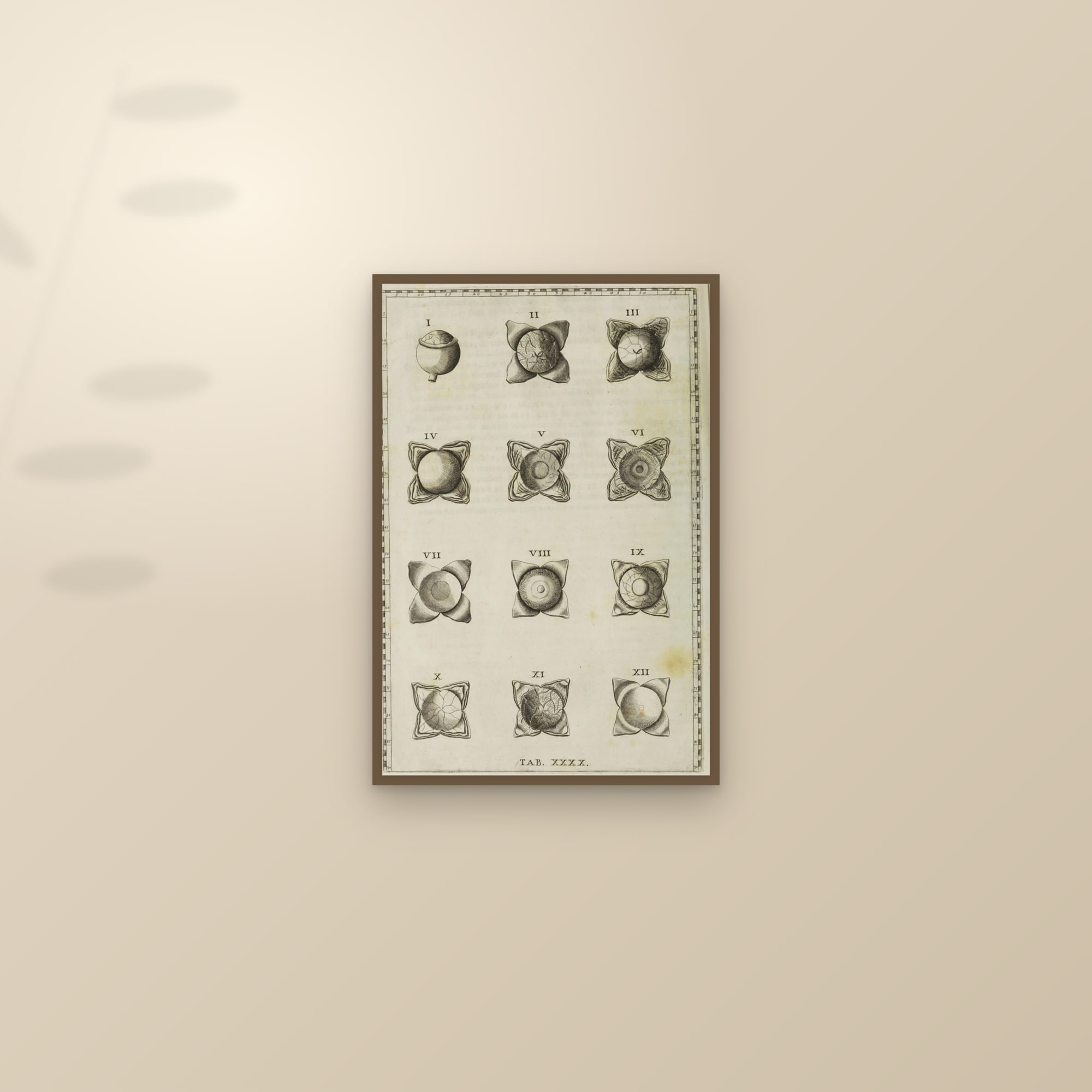
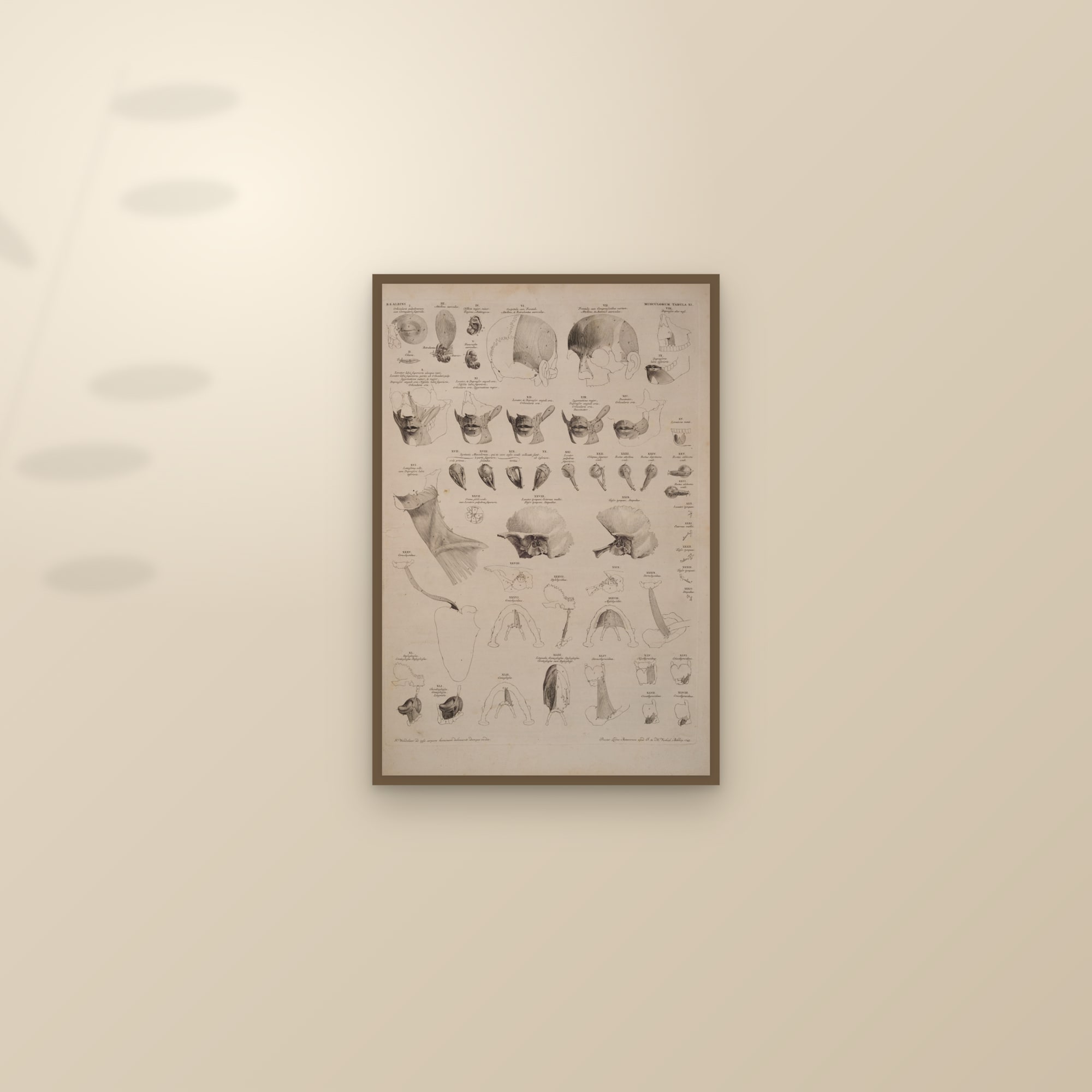
The eye and the ear from the atlases: Johannes Sobotta's colour plates of 1920, the eye in horizontal section, the retina, the eyeball and its muscles, the orbit, the eye in the orbit, the blood vessels of the eye and the eye with its lacrimal apparatus; four plates of the ear, the inner ear, the bony labyrinth, the cochlea and semicircular canals and the middle ear in section; and Bartolomeo Eustachi's engraving of the eye in twelve dissections, published in 1714.
12 prints · printed to order on heavyweight matte paper · framed or unframed
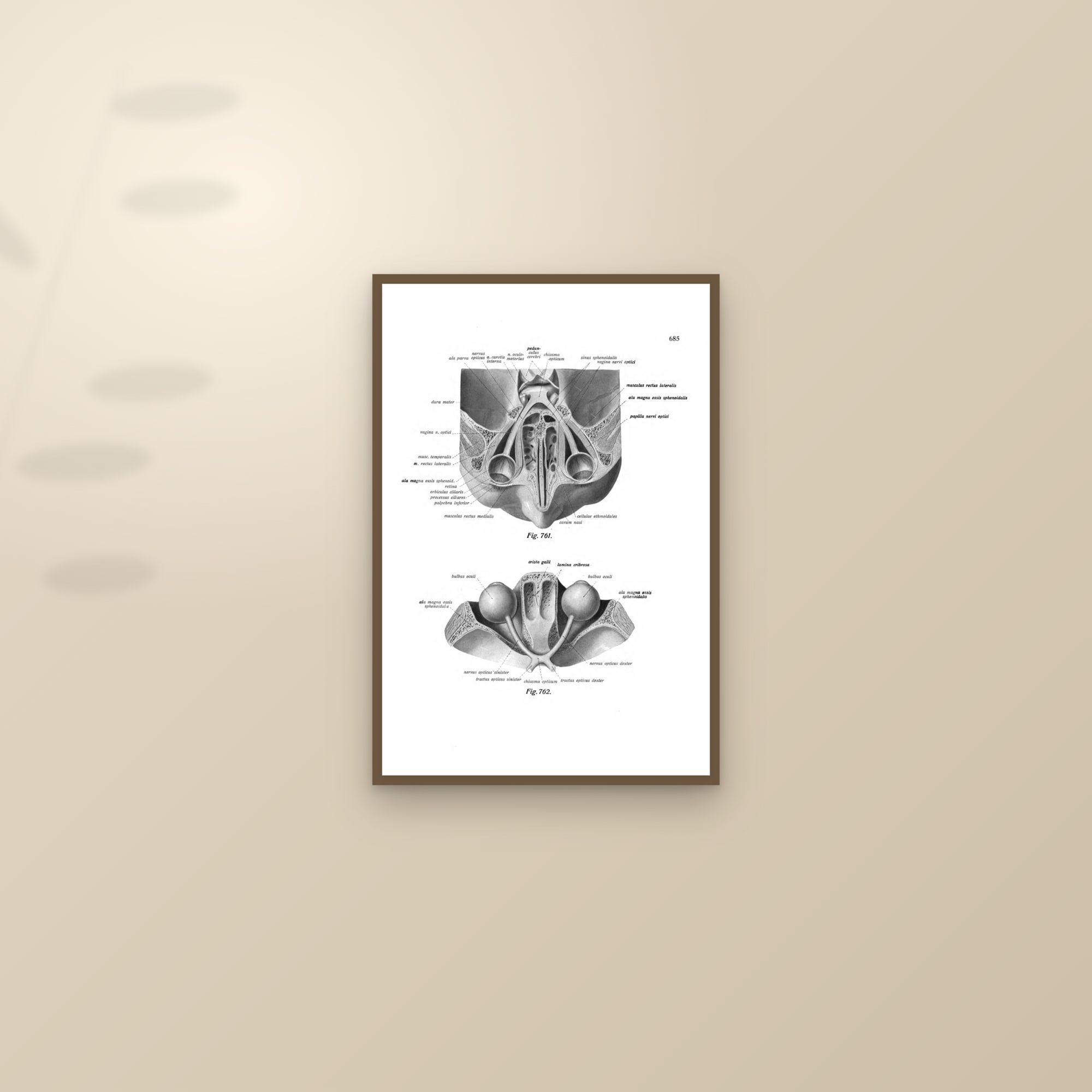
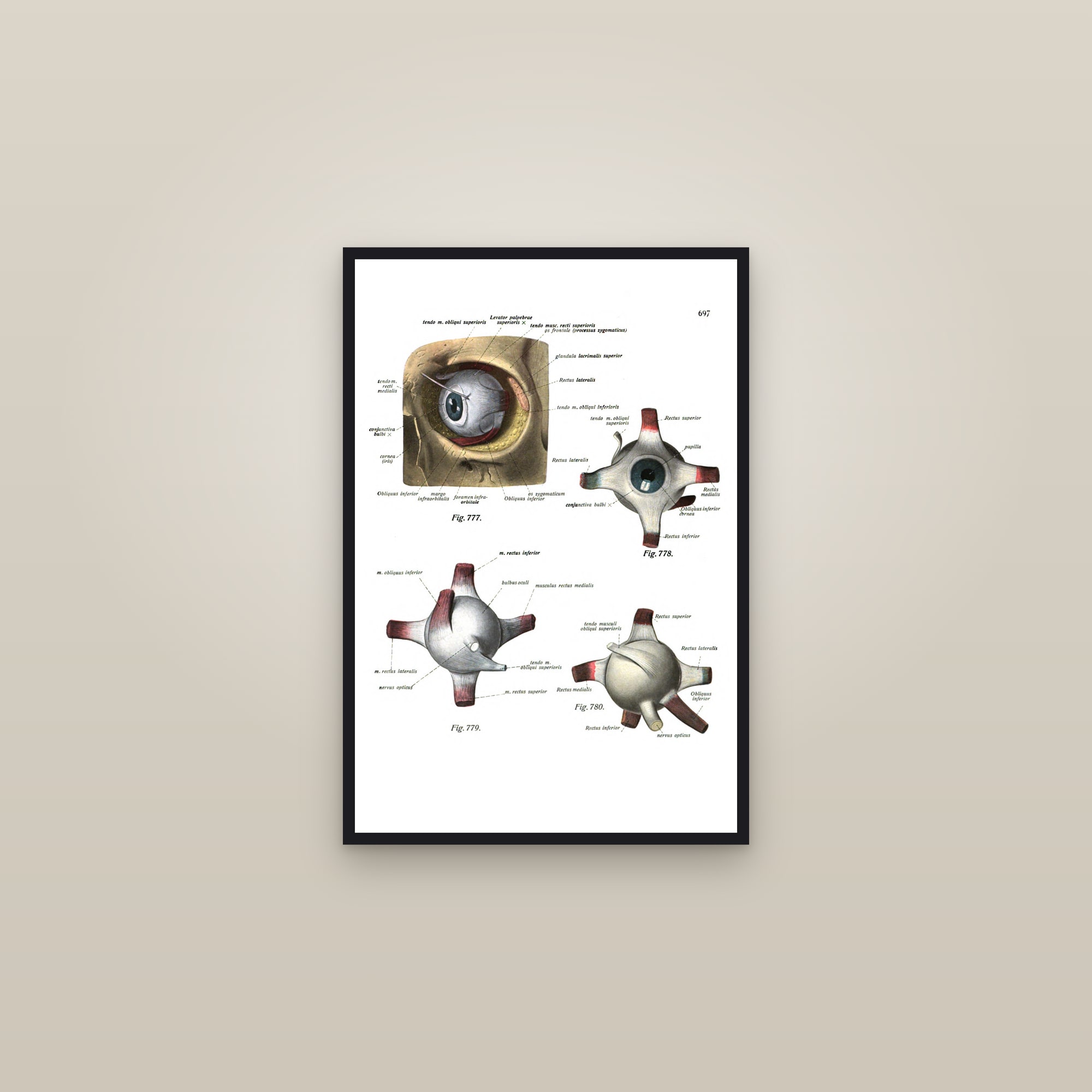
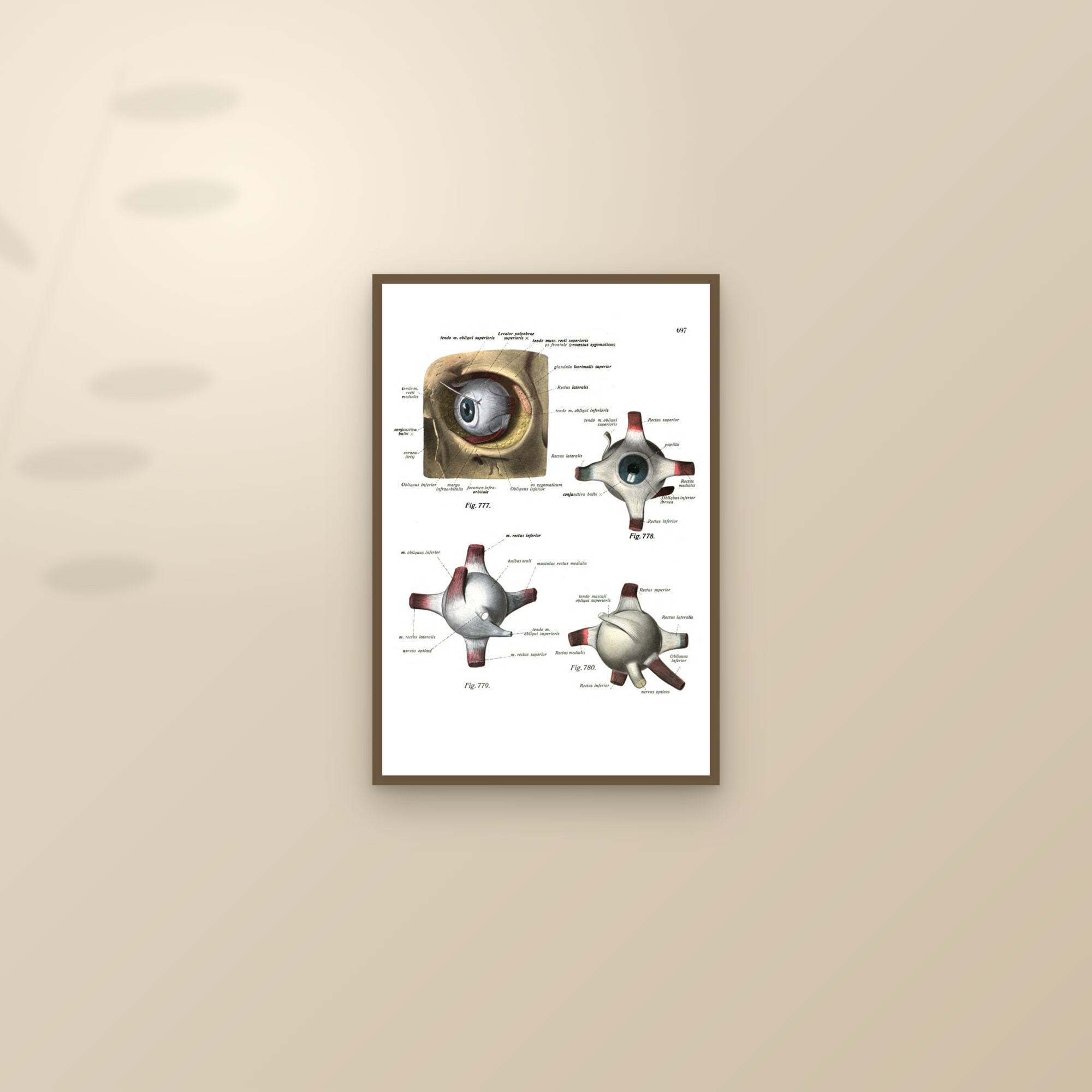
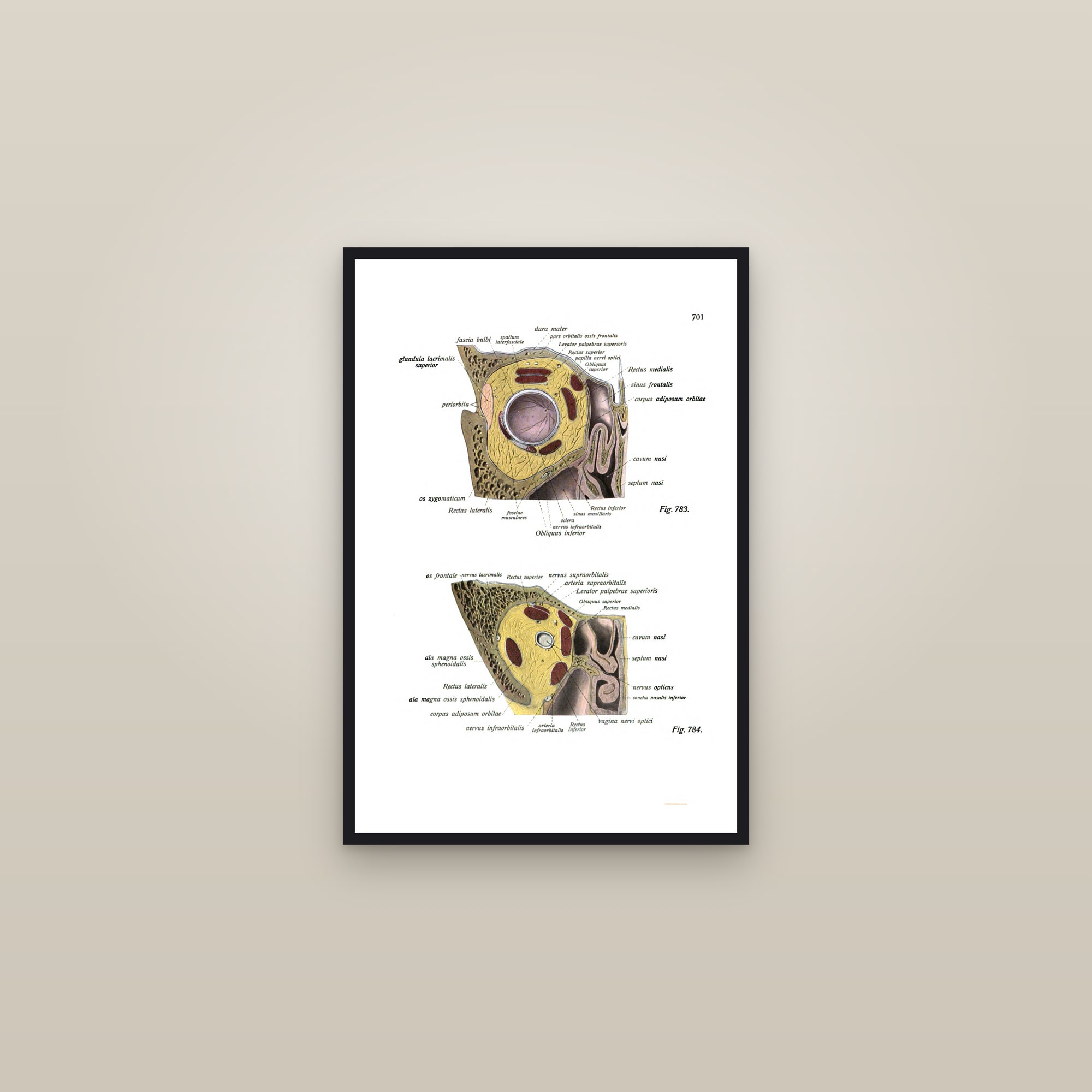
Sobotta's eye
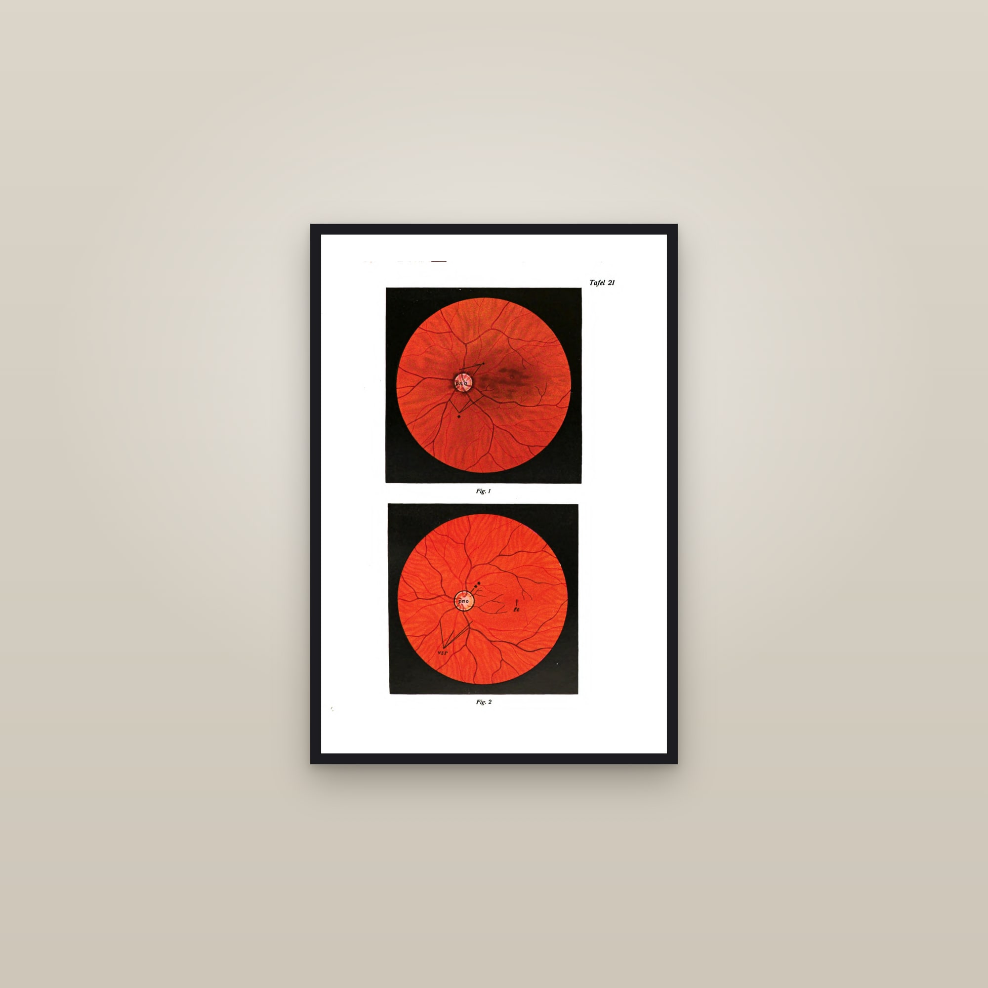
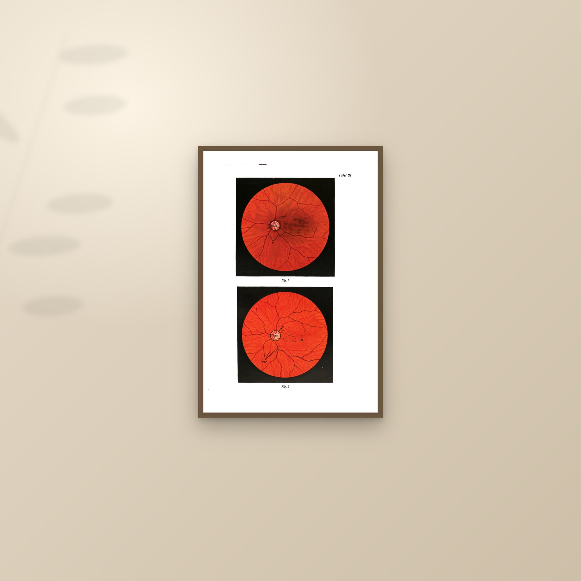
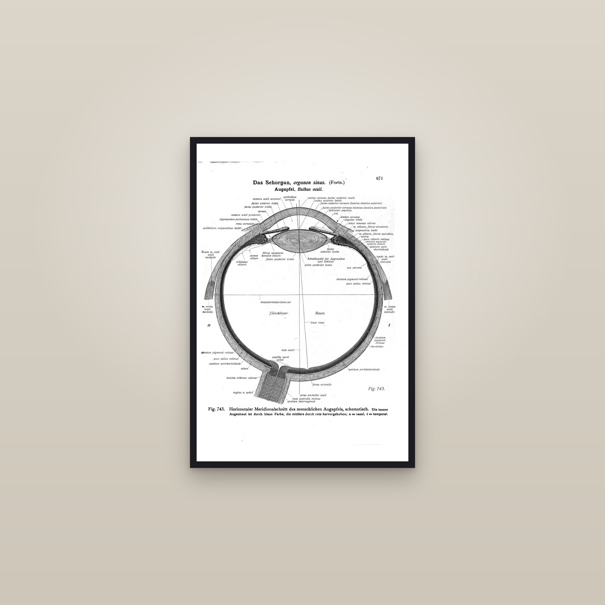
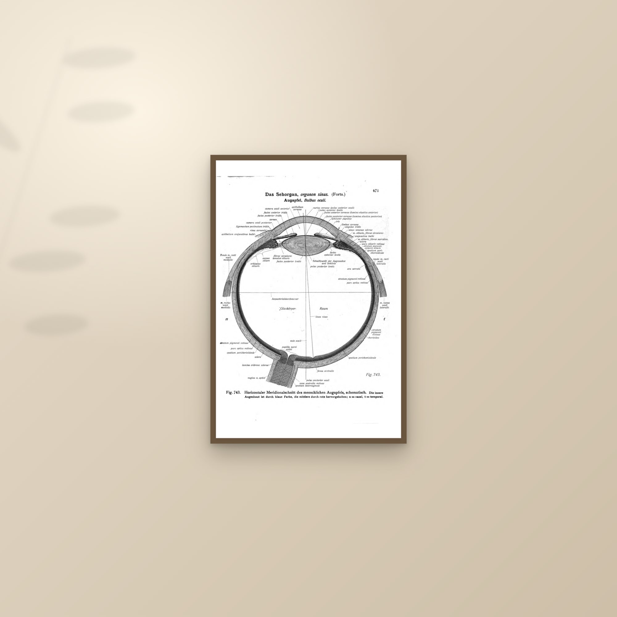
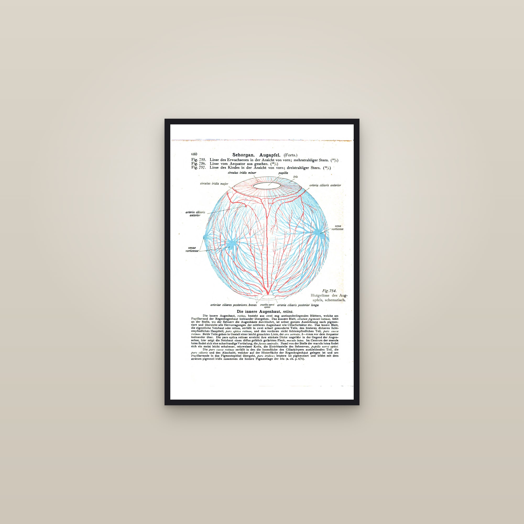
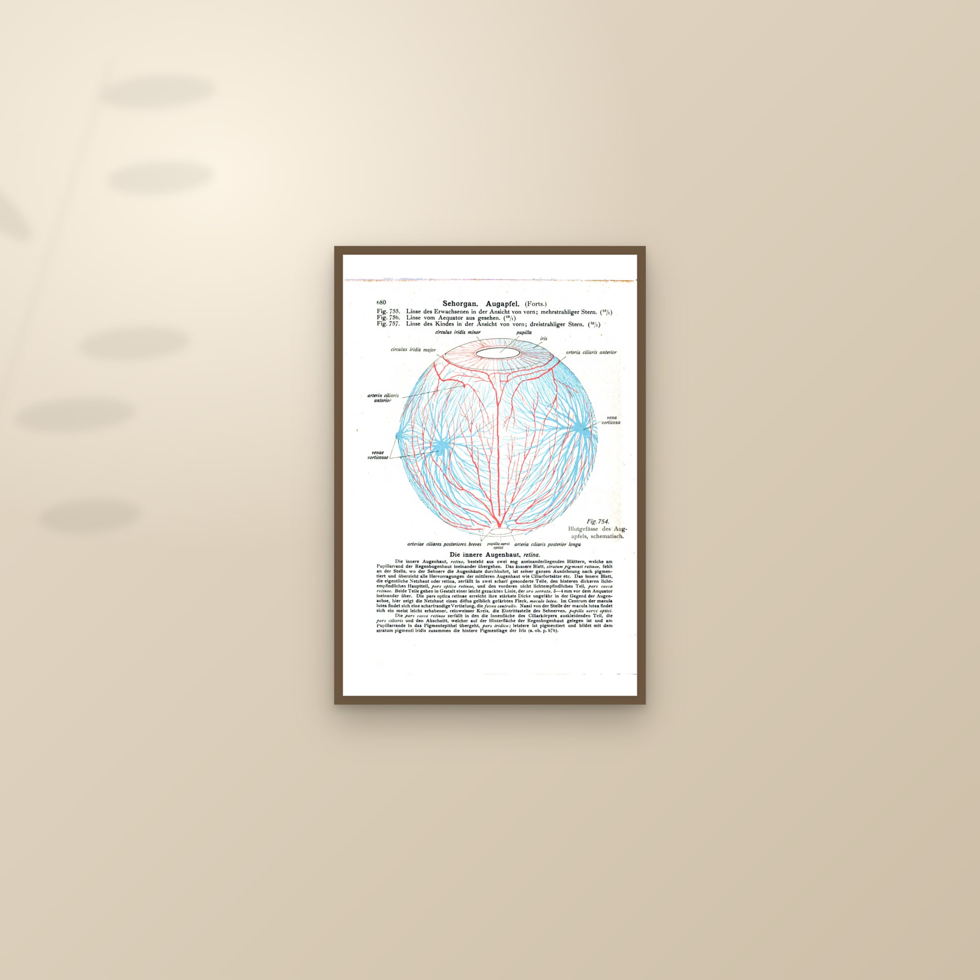
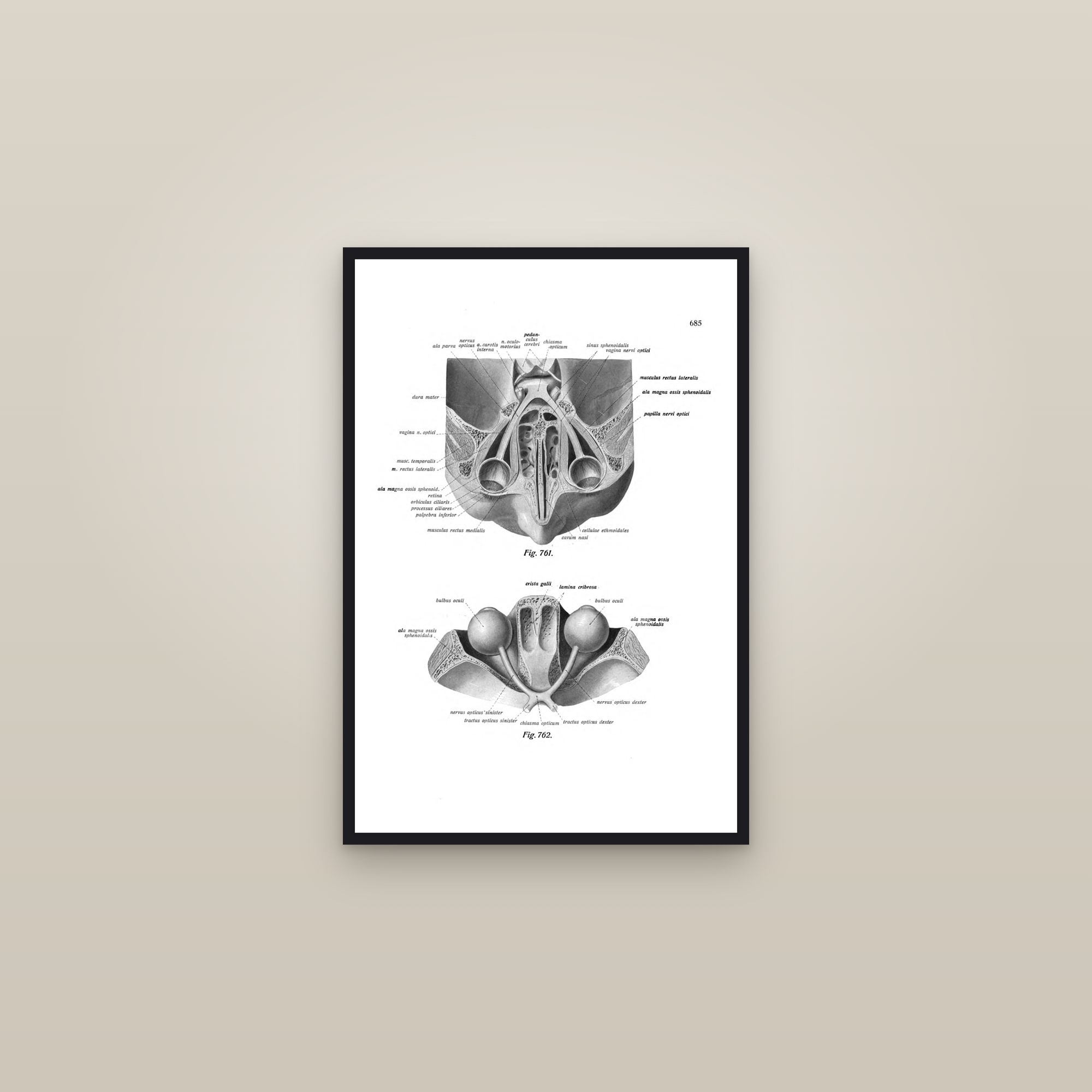
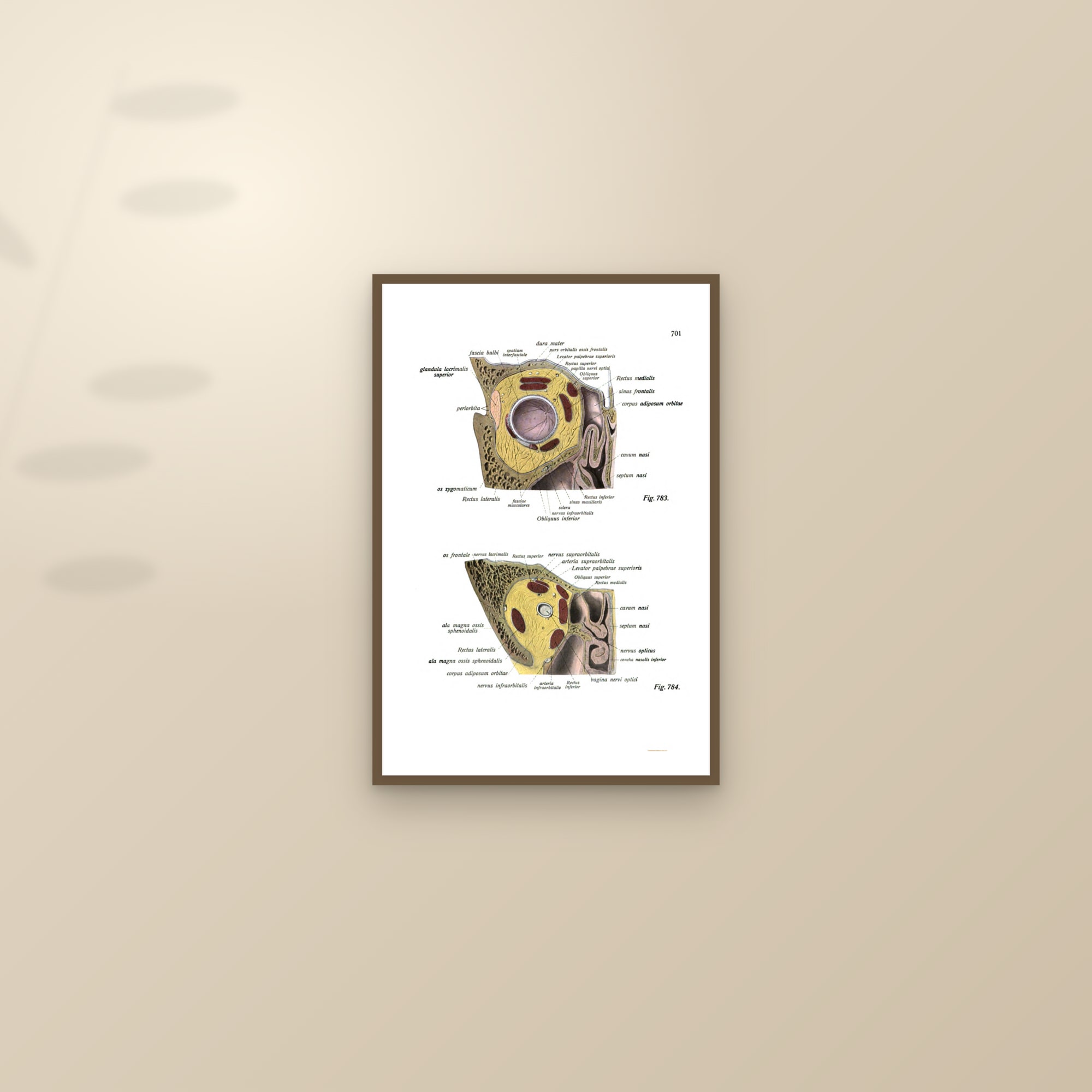
Seven Sobotta plates cover the eye: the eye in horizontal section, the plate that shows cornea, lens, vitreous and retina in one cut; the retina itself, the fundus as seen through an ophthalmoscope; the eyeball with its six muscles; the orbit and the eye muscles; the eye sitting in the orbit; the blood vessels of the eye; and the eye with its lacrimal apparatus in section. They are chromolithographs from the third volume of the atlas, 1920, and the horizontal section is the plate every optometrist recognises.
The ear
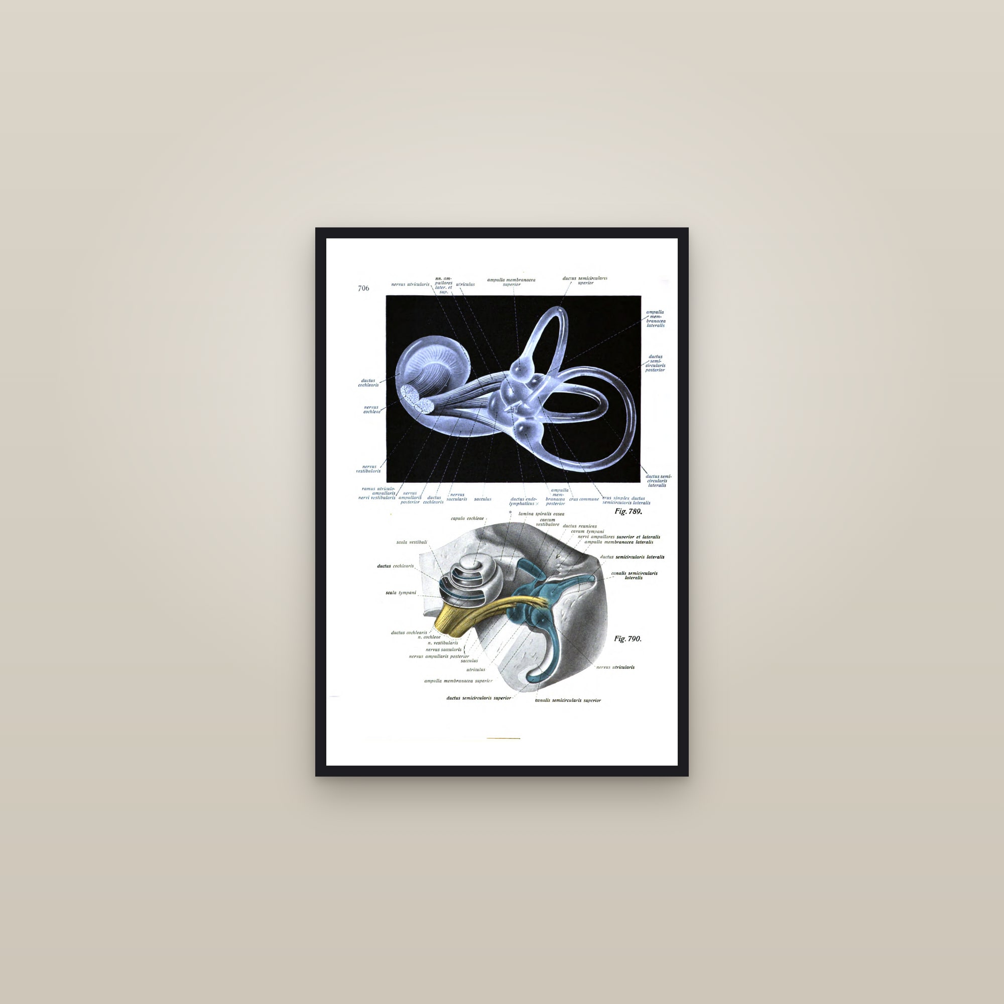
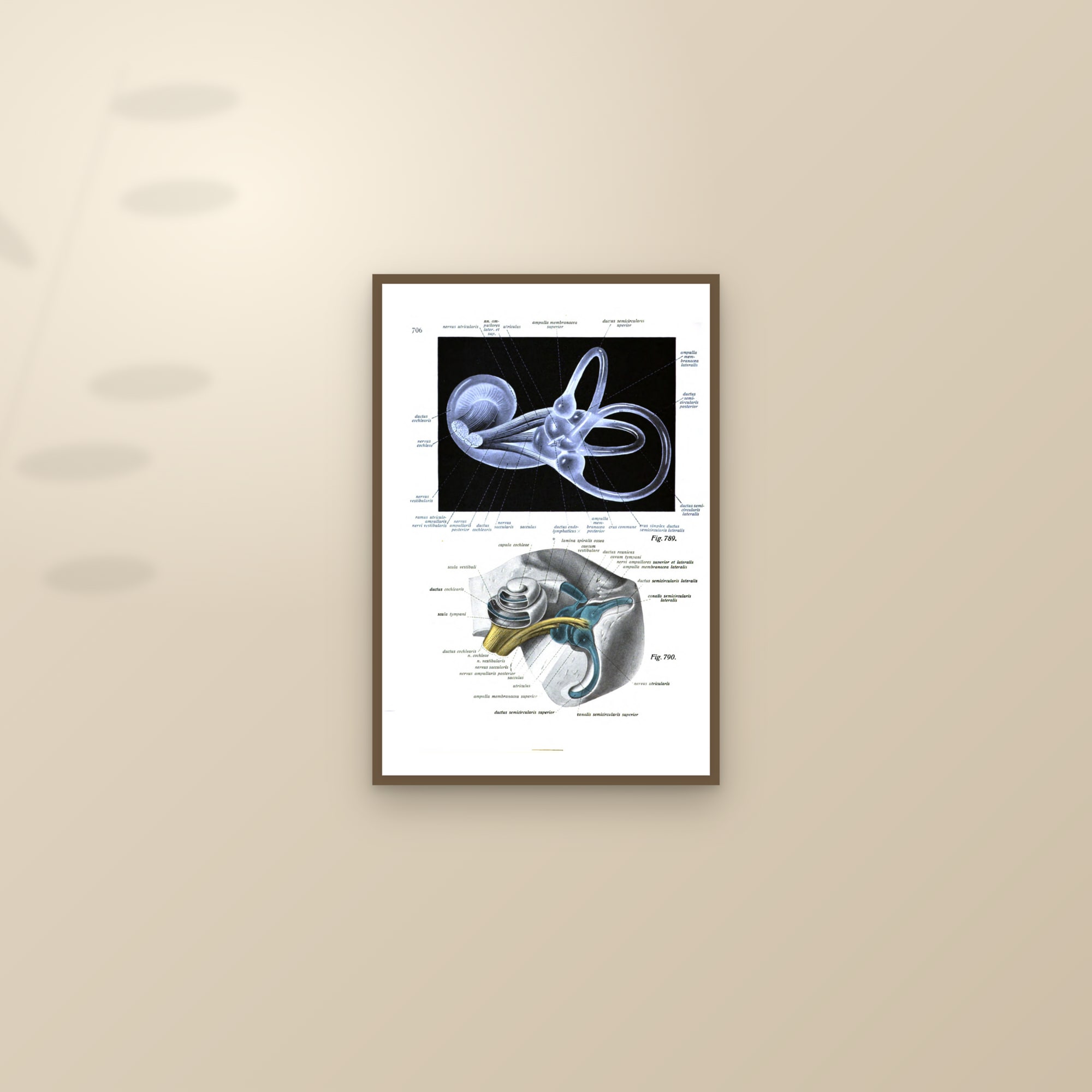
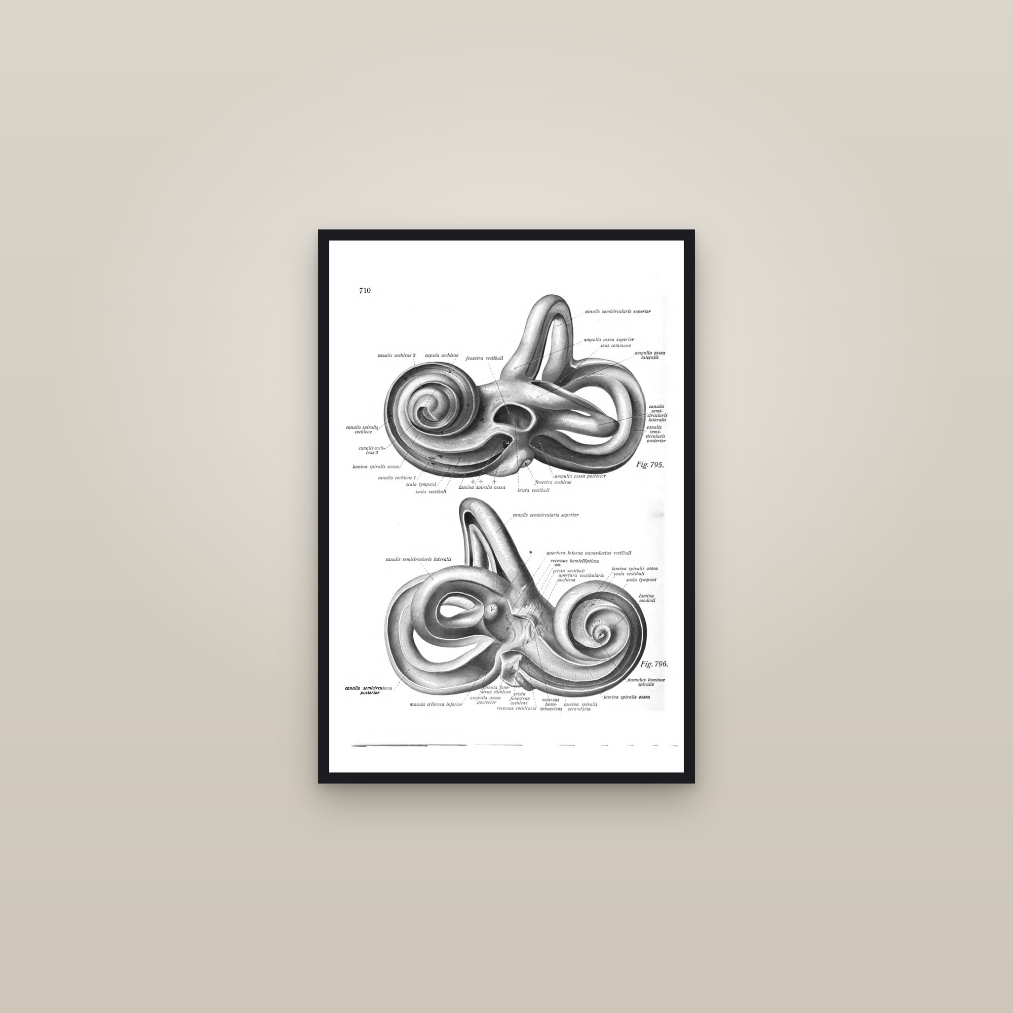
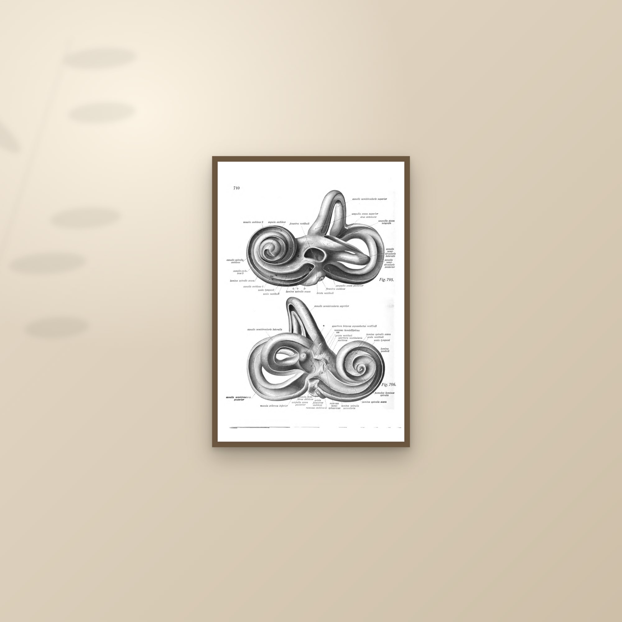
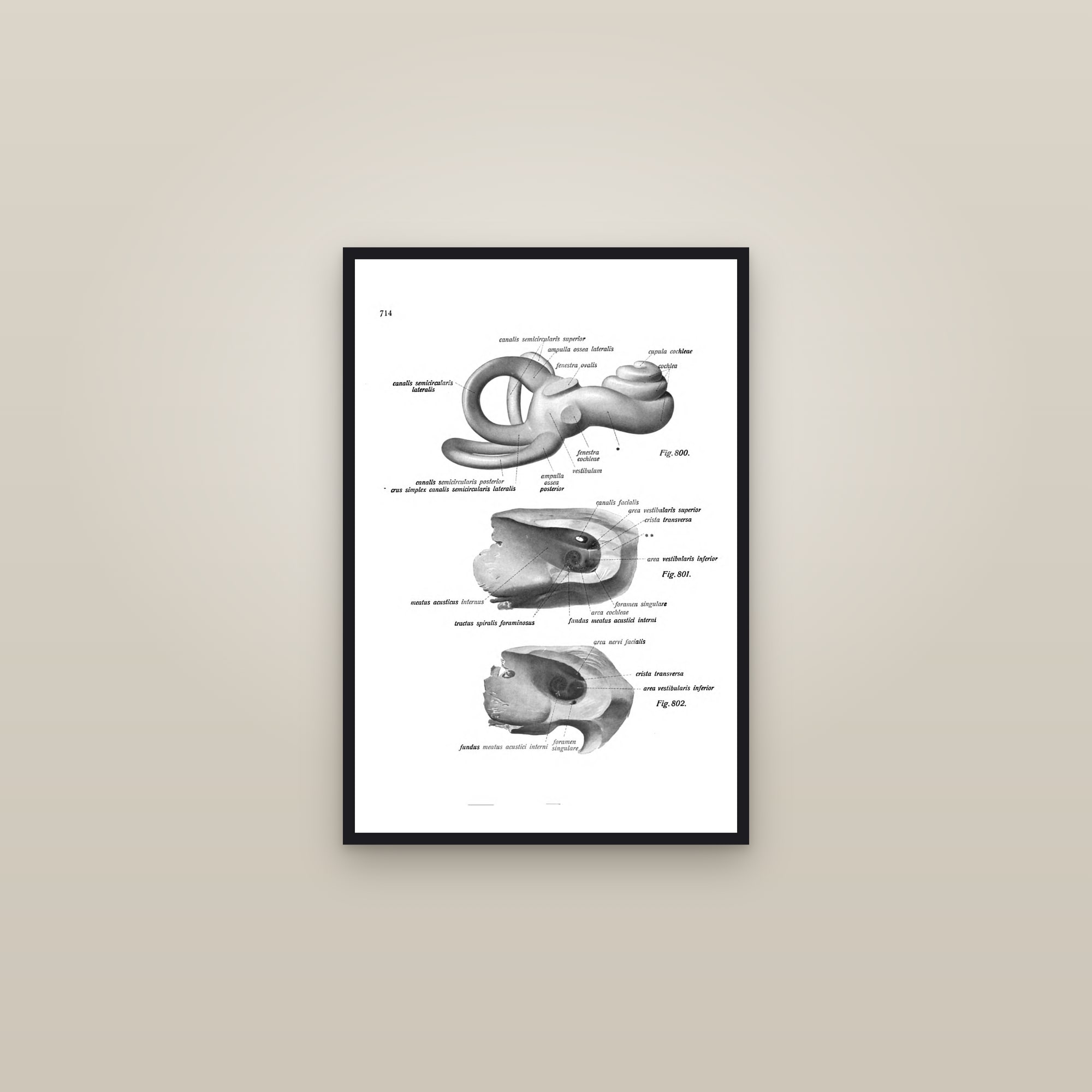
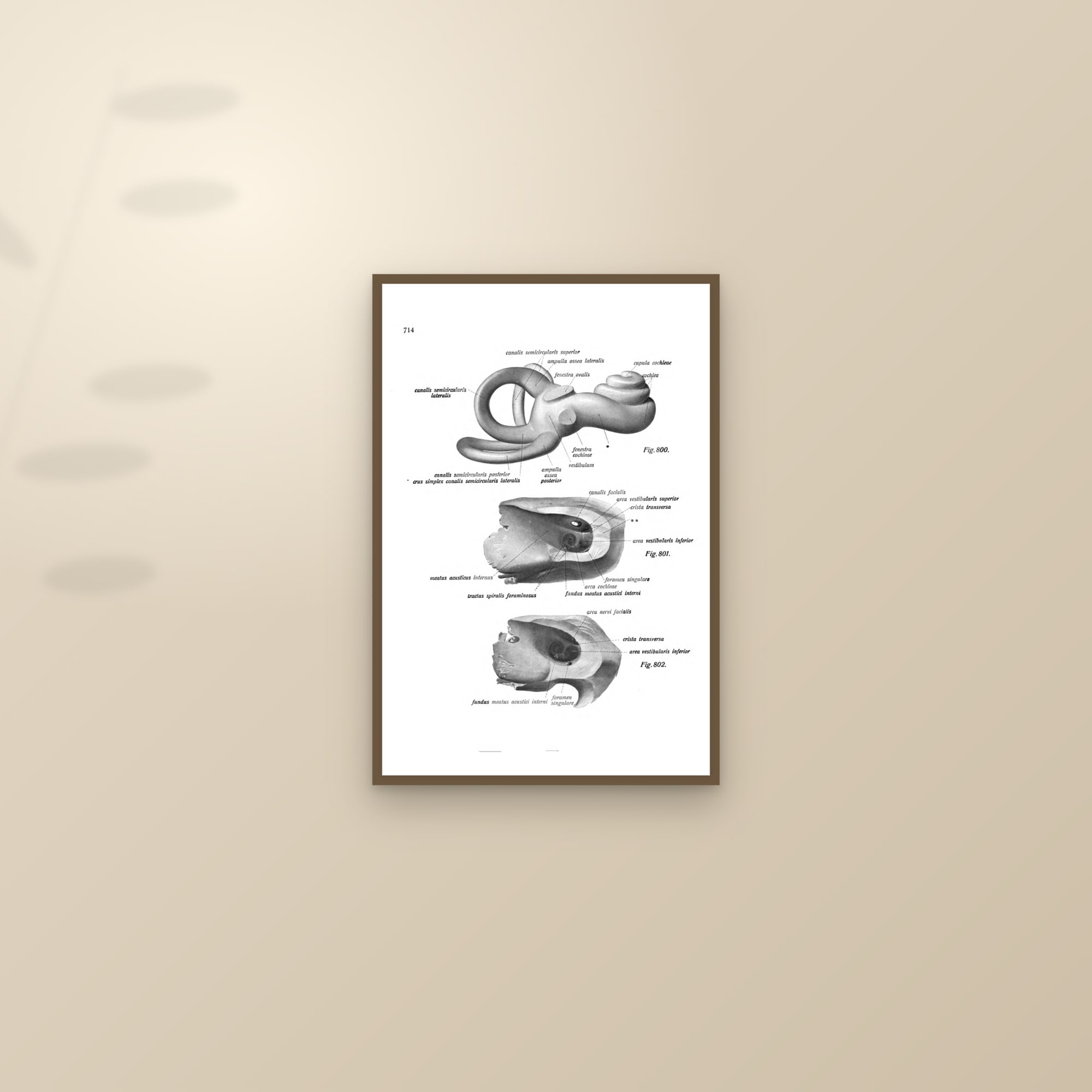
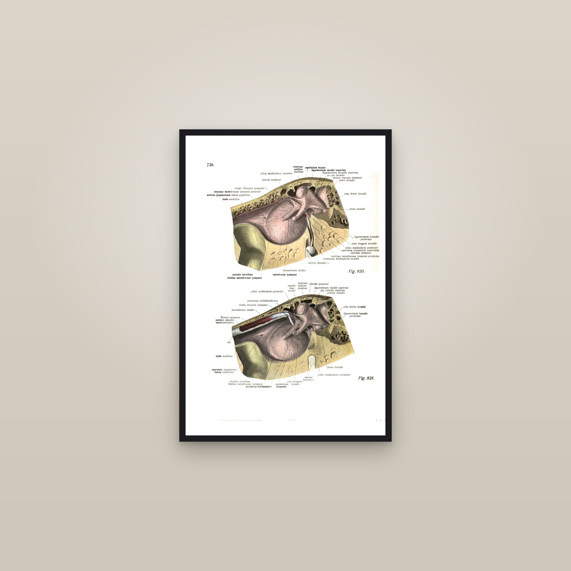
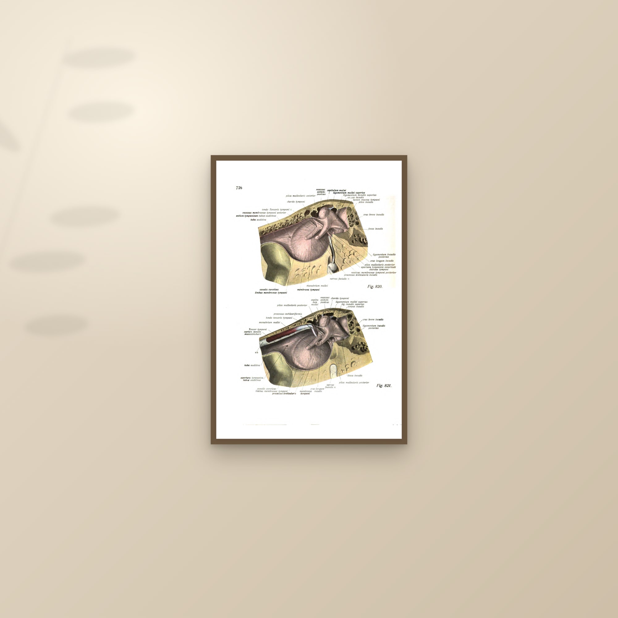
Four plates from the same volume take the ear: the inner ear with its labyrinth printed in colour, the bony labyrinth on its own, the cochlea with the semicircular canals, and the middle ear in section with the ossicles. The structures are small and the drawings are correspondingly fine, so these hang best at 16×24 where the detail can be read, and they suit the ENT consulting room and the audiologist's office. Eustachi, who gave his name to the Eustachian tube, supplies the historical anchor for the set with his twelve dissections of the eye.
Where they hang
Optometrists, ophthalmologists and opticians hang the eye plates in the consulting room and behind the dispensing desk; ENT surgeons and audiologists take the ear. At home they go in the study, and the retina plate, a warm orange disc on a pale ground, is the one that works as plain abstract art in a hallway. Sobotta plates print up to 24×36, the Eustachi engraving up to 16×24. Each is offered full-bleed or as a captioned specimen on cream, unframed or framed in black or dark wood with no mat.