Foot Anatomy Prints
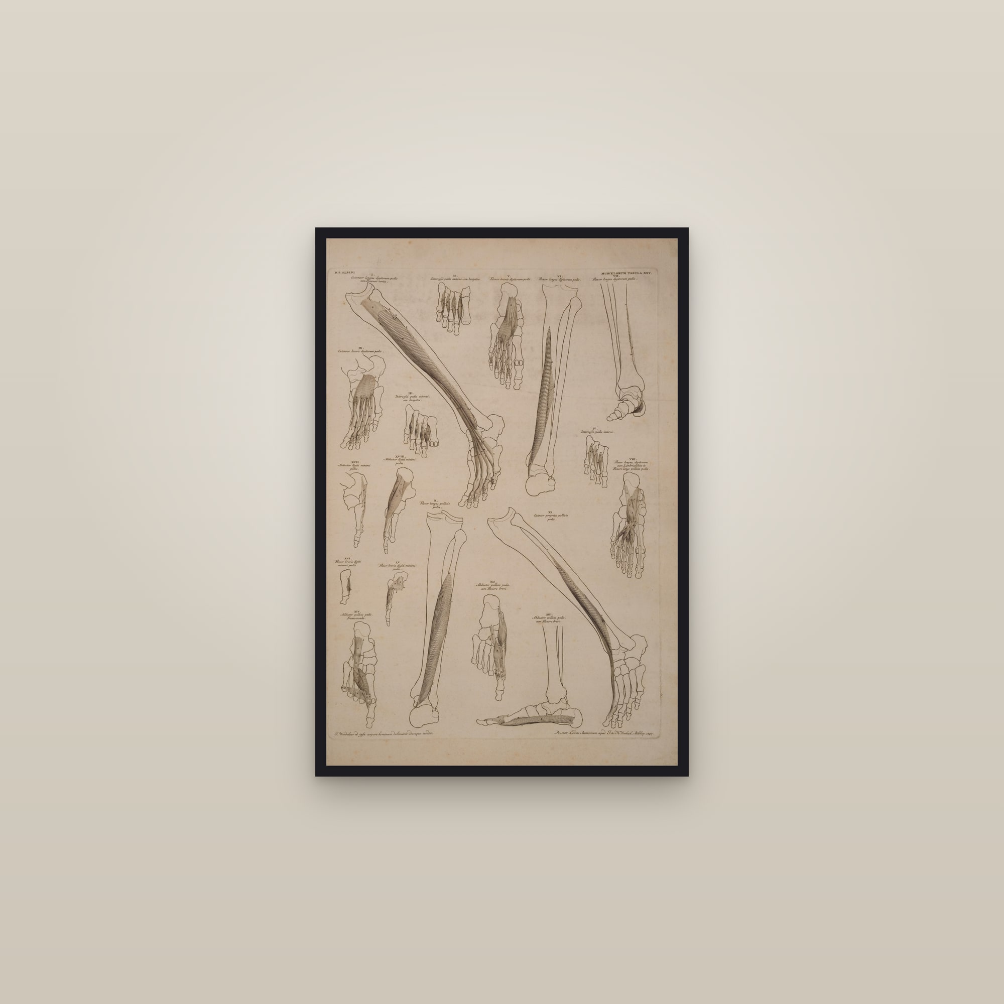
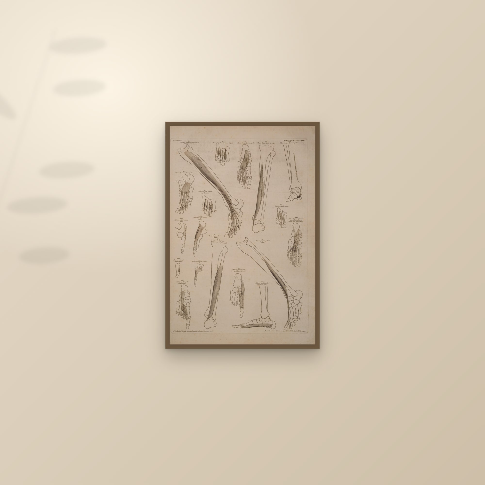
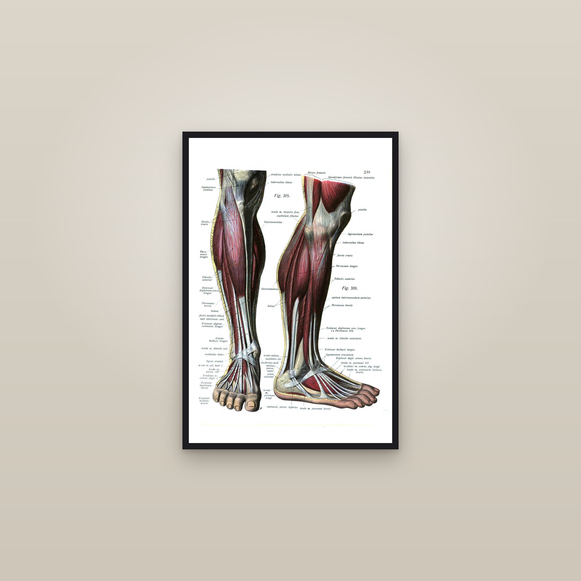
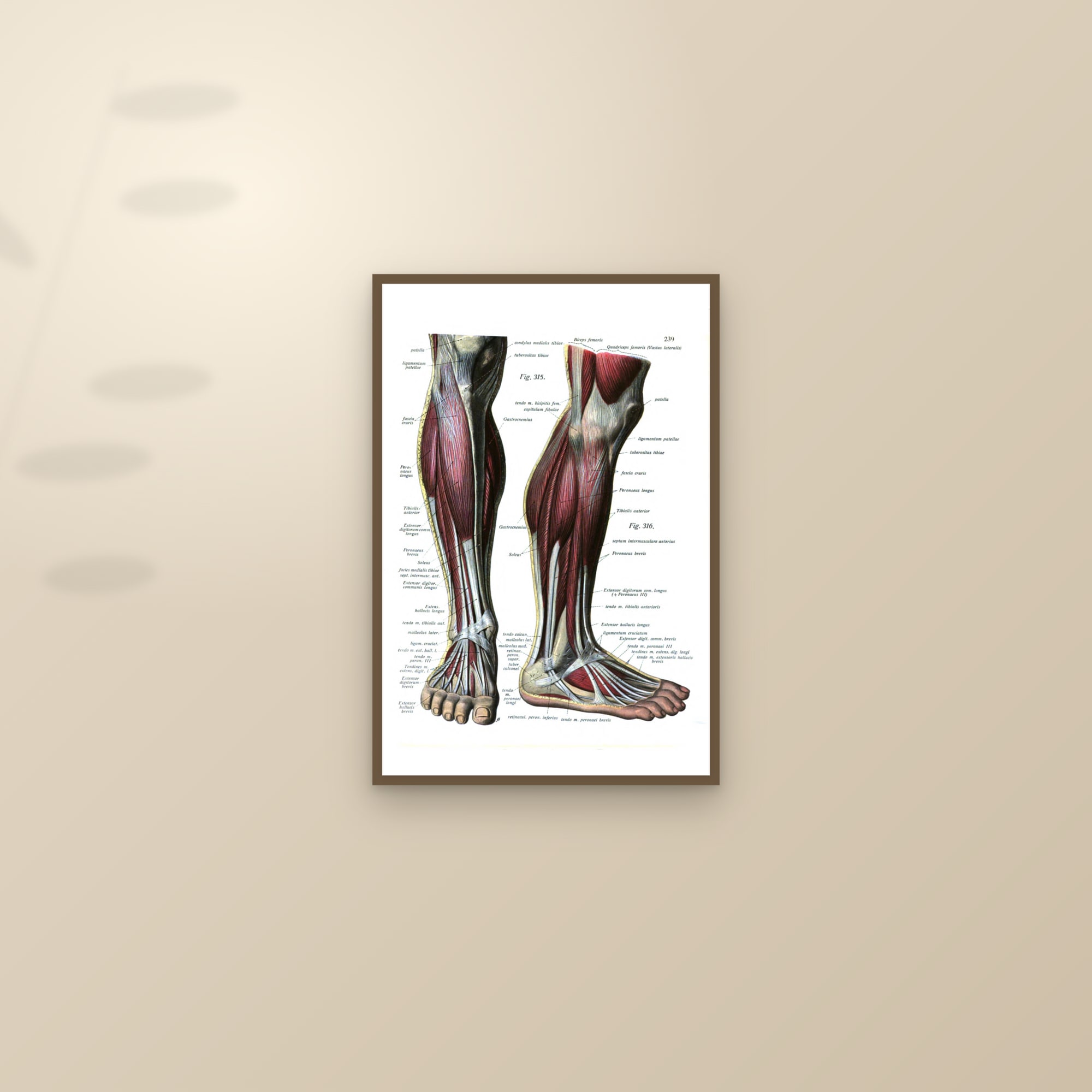
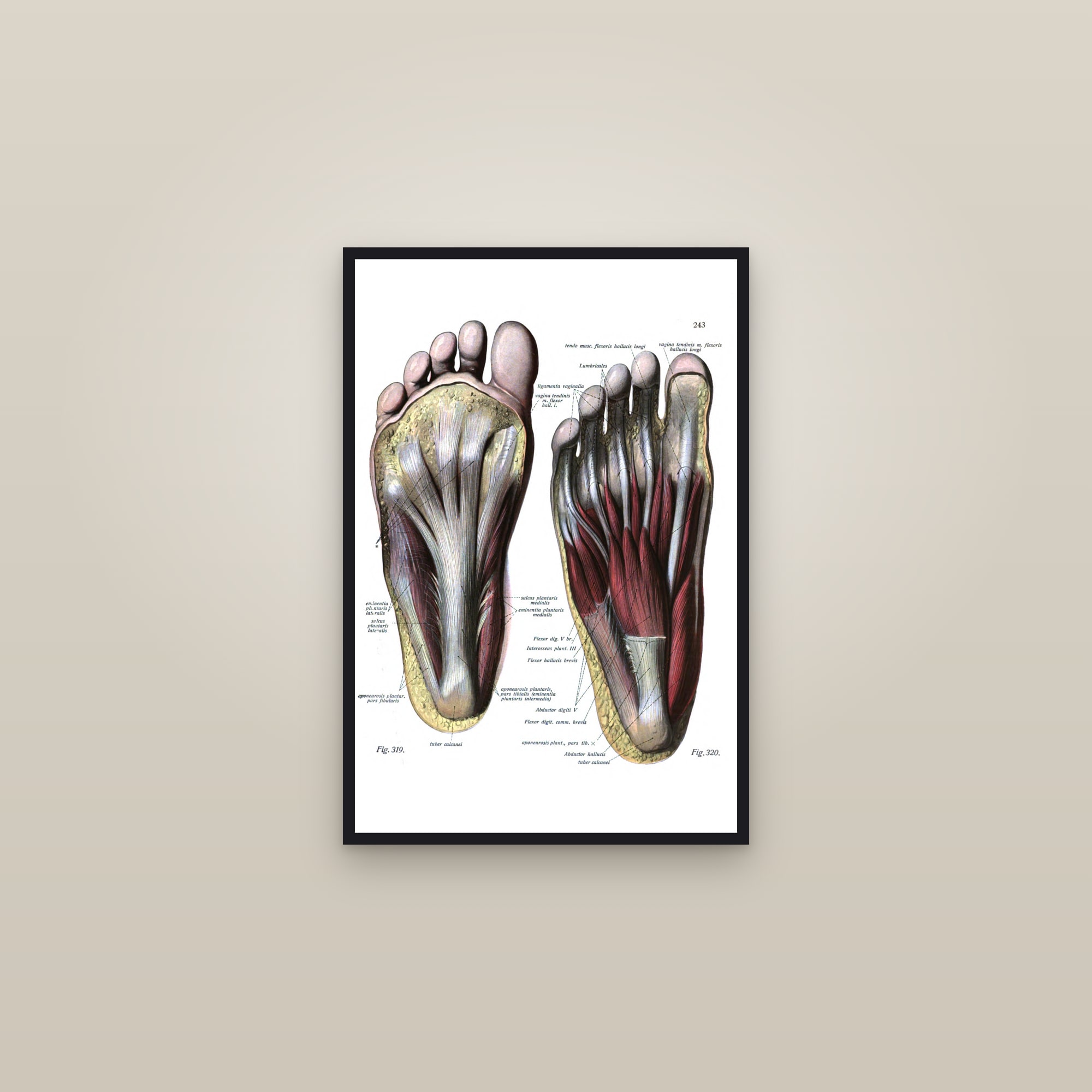
The foot and ankle from the atlases: Johannes Sobotta's colour plates of 1919, the bones of the foot from above and from below and the side, the muscles of the lower leg and foot, the muscles of the sole in a superficial and a deep layer, and the ligaments of the foot and ankle; with Albinus's engravings of the muscles of the foot and lower leg from the Tabulae sceleti et musculorum of 1747.
6 prints · printed to order on heavyweight matte paper · framed or unframed
The plates
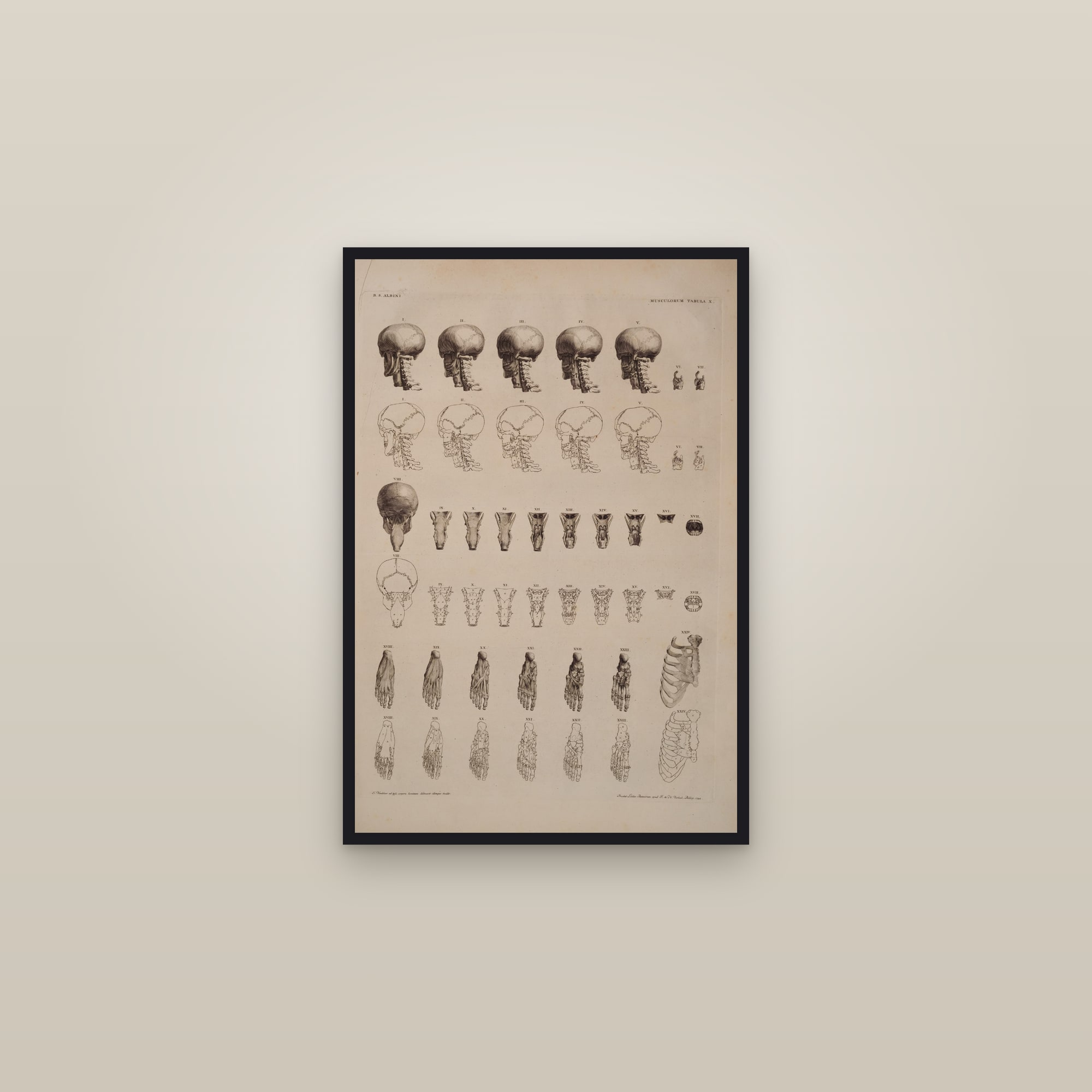
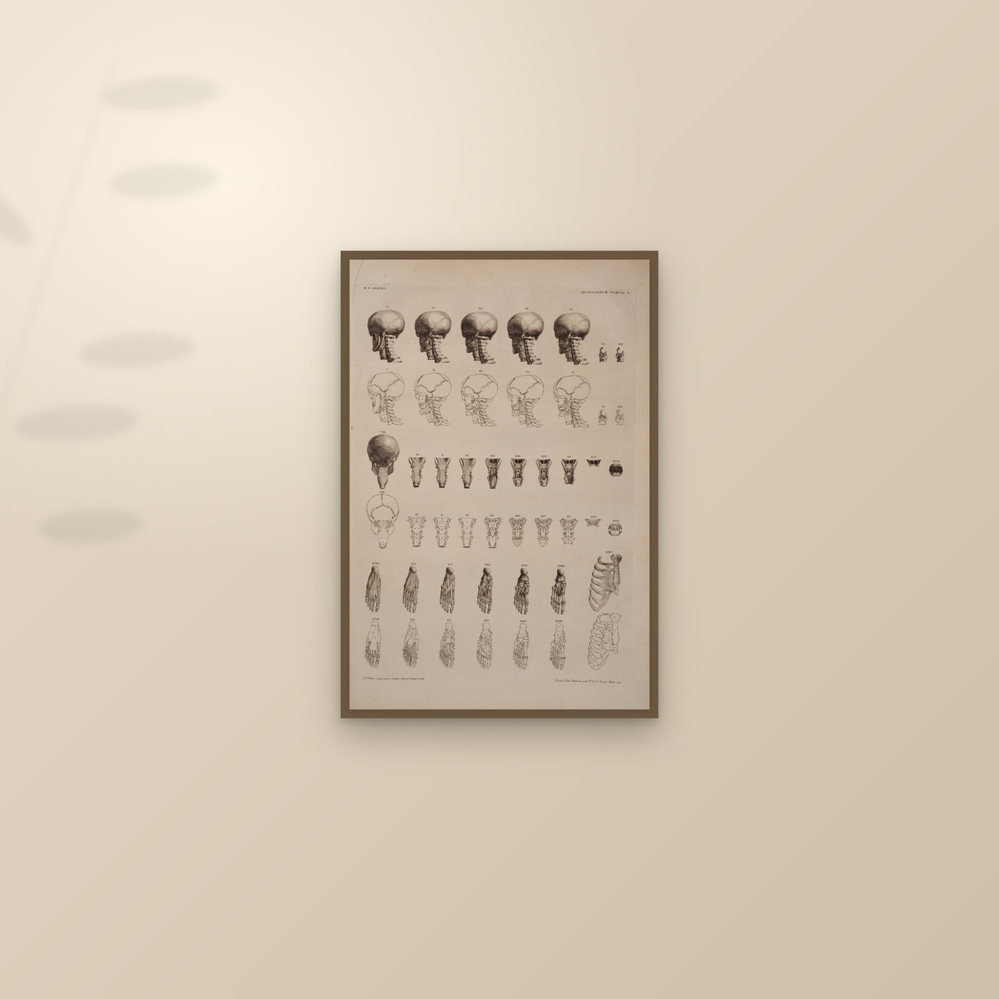
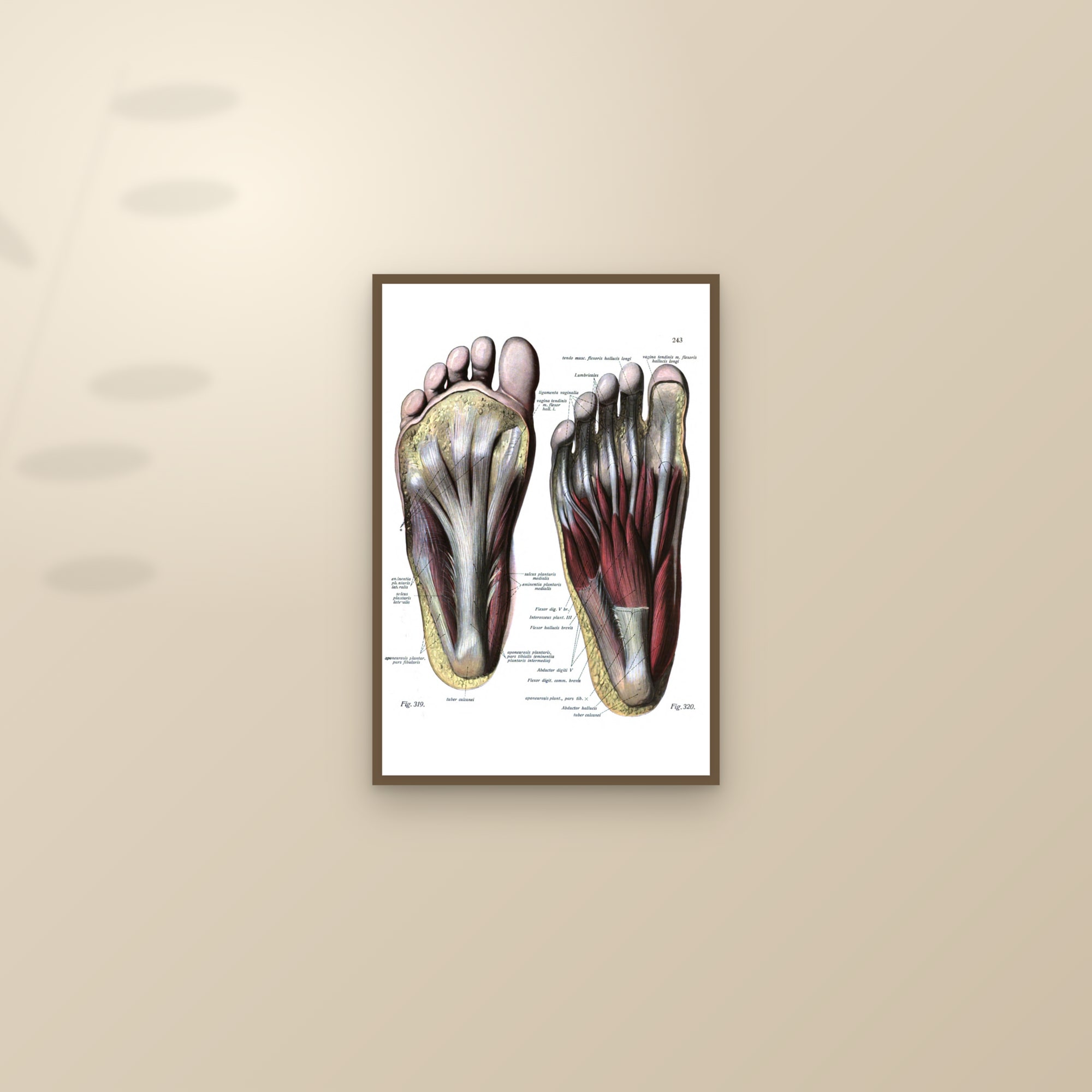
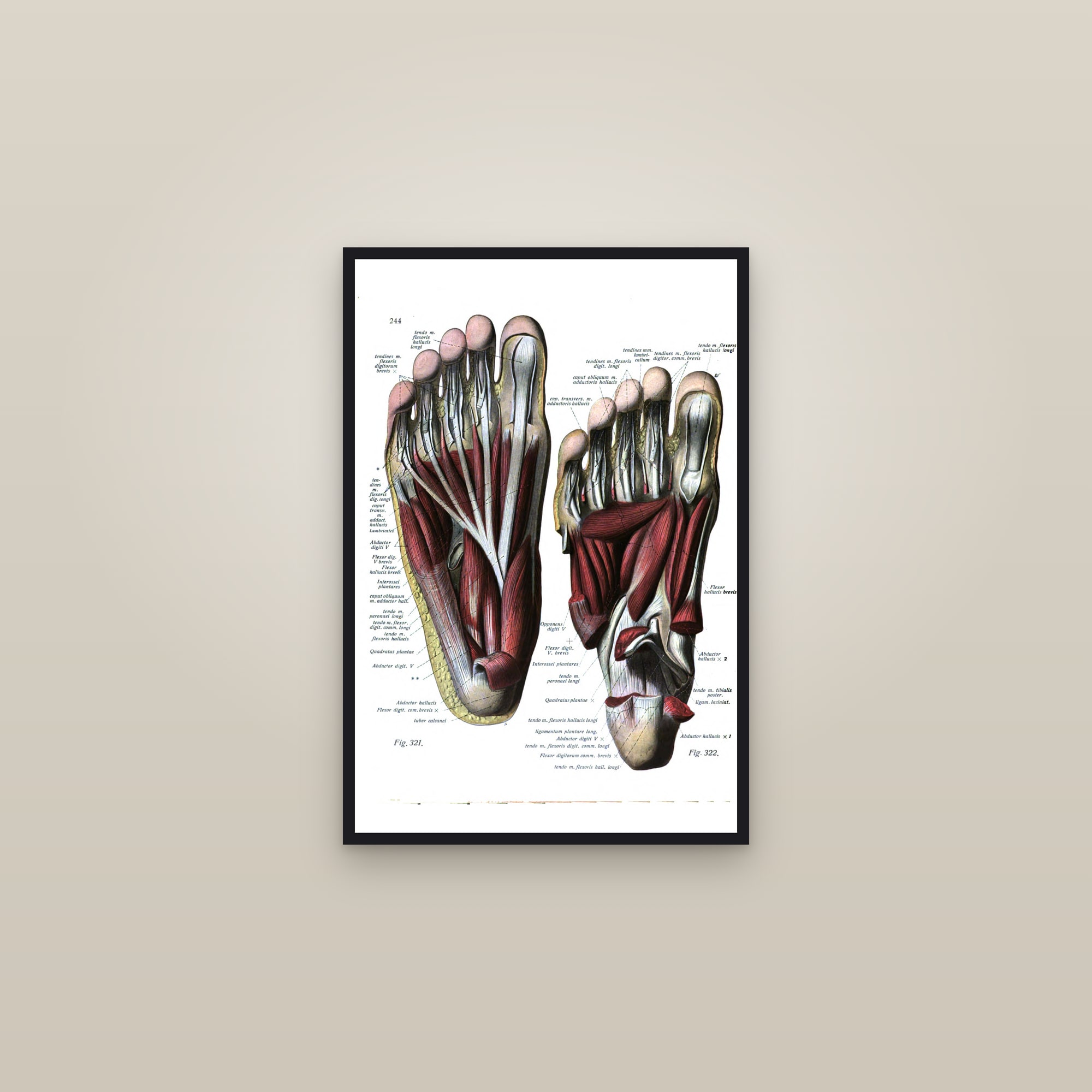
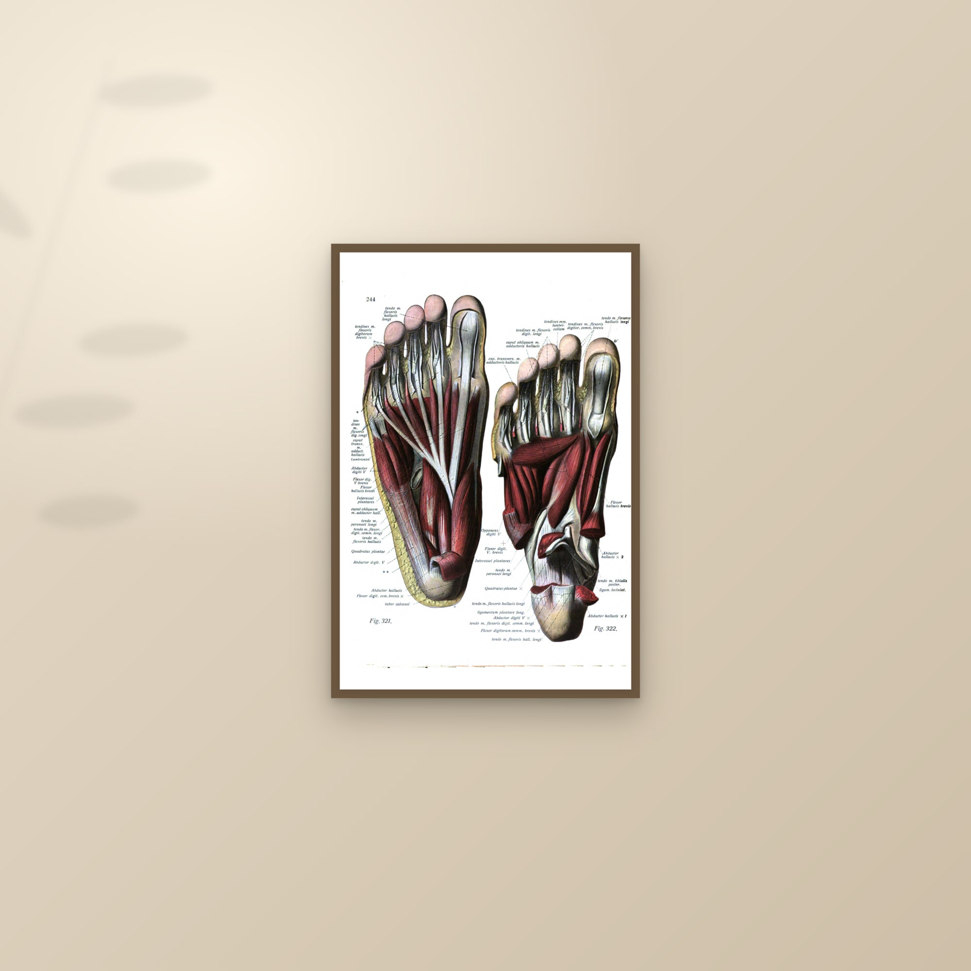
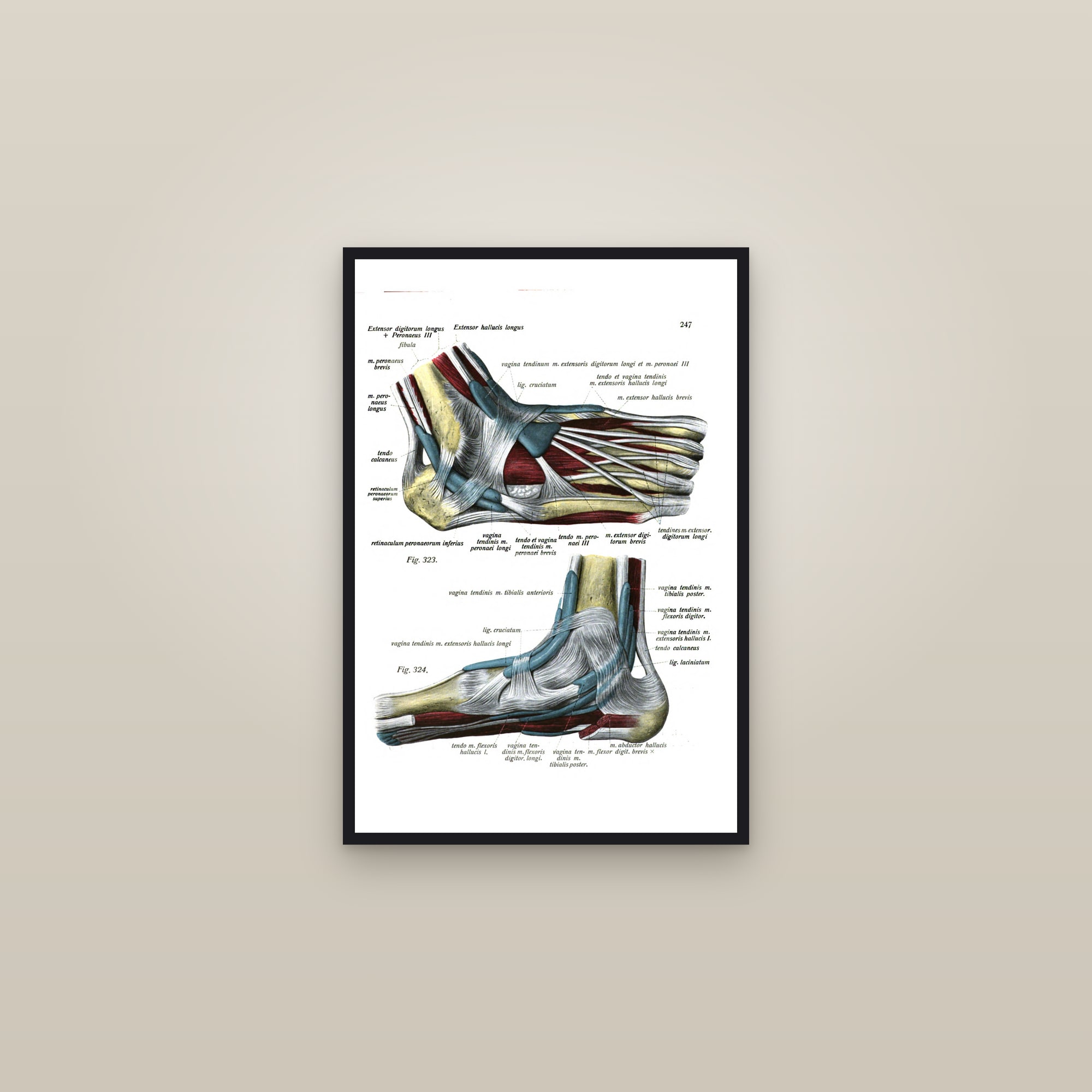
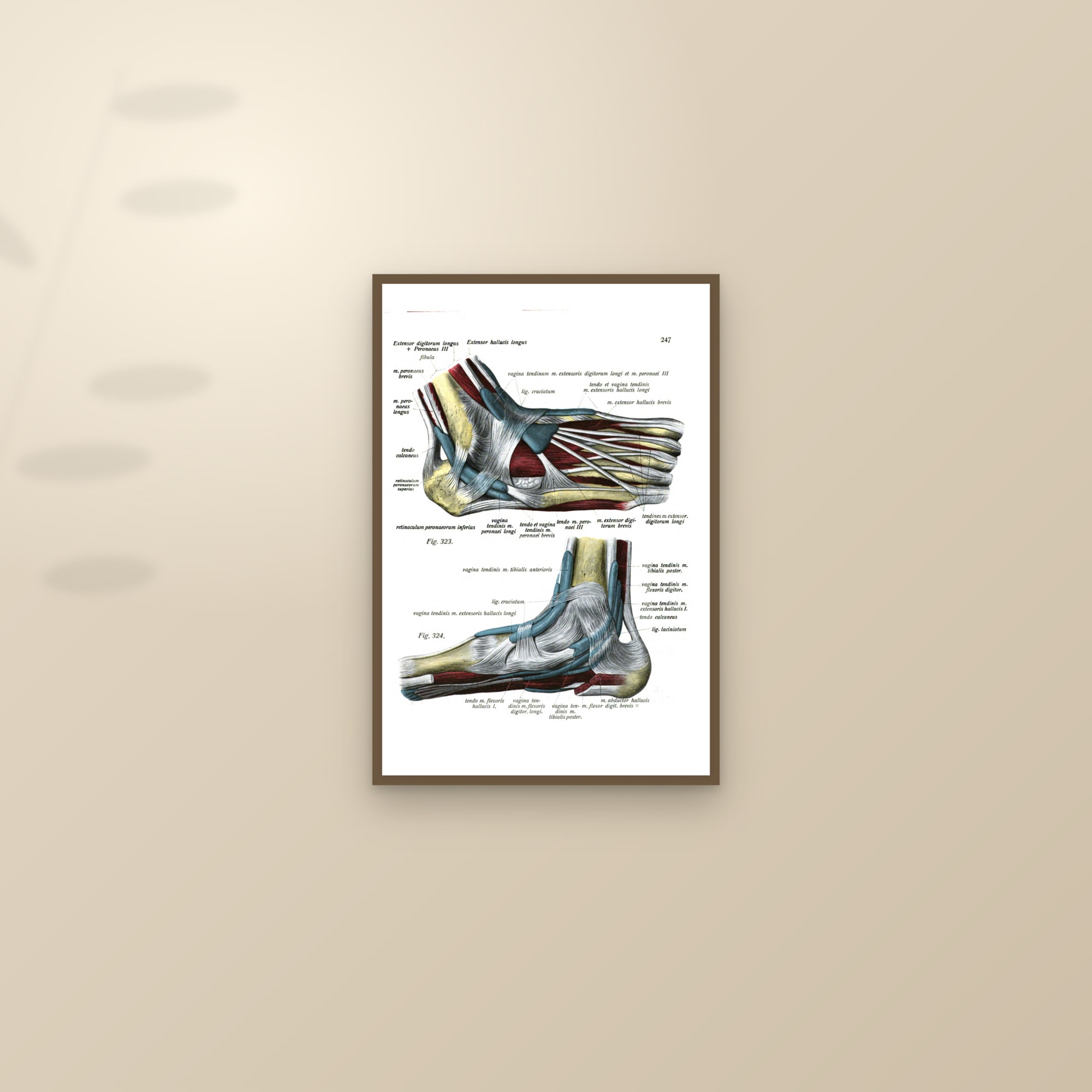
Six Sobotta chromolithographs from the 1919 atlas cover the foot from the bones out: the bones of the foot seen from above, the same bones from below and from the side, the muscles of the lower leg and foot in side view, the muscles of the sole of the foot and the deep muscles beneath them, and the ligaments of the foot and ankle. Albinus adds two engraved study plates of 1747, the muscles of the foot and lower leg and a study sheet of the neck, larynx and foot. The bones-from-above plate, a single foot on a pale ground, is the one most often hung alone.
For the podiatrist, the physio and the runner
Podiatrists and chiropodists hang these in the treatment room; physiotherapists and sports therapists take the ligaments of the ankle, the plate that explains most of their caseload; runners, dancers and climbers hang the bones of the foot in the home gym or the study. The ligaments plate at 24×36 is clear from across a room; the sole plates read better at 16×24.
Sizes and framing
Every plate on this page prints up to 24×36 (the Sobotta and Albinus scans support the full ladder), and each is offered full-bleed or as a captioned specimen on cream with its title, atlas and date typeset beneath, on 250 gsm off-white archival paper. Frames are black or dark wood with a plexiglass front and no mat. Bones from above and bones from below hang as a pair; add the ligaments for a trio and three or more prints take 10% off automatically.