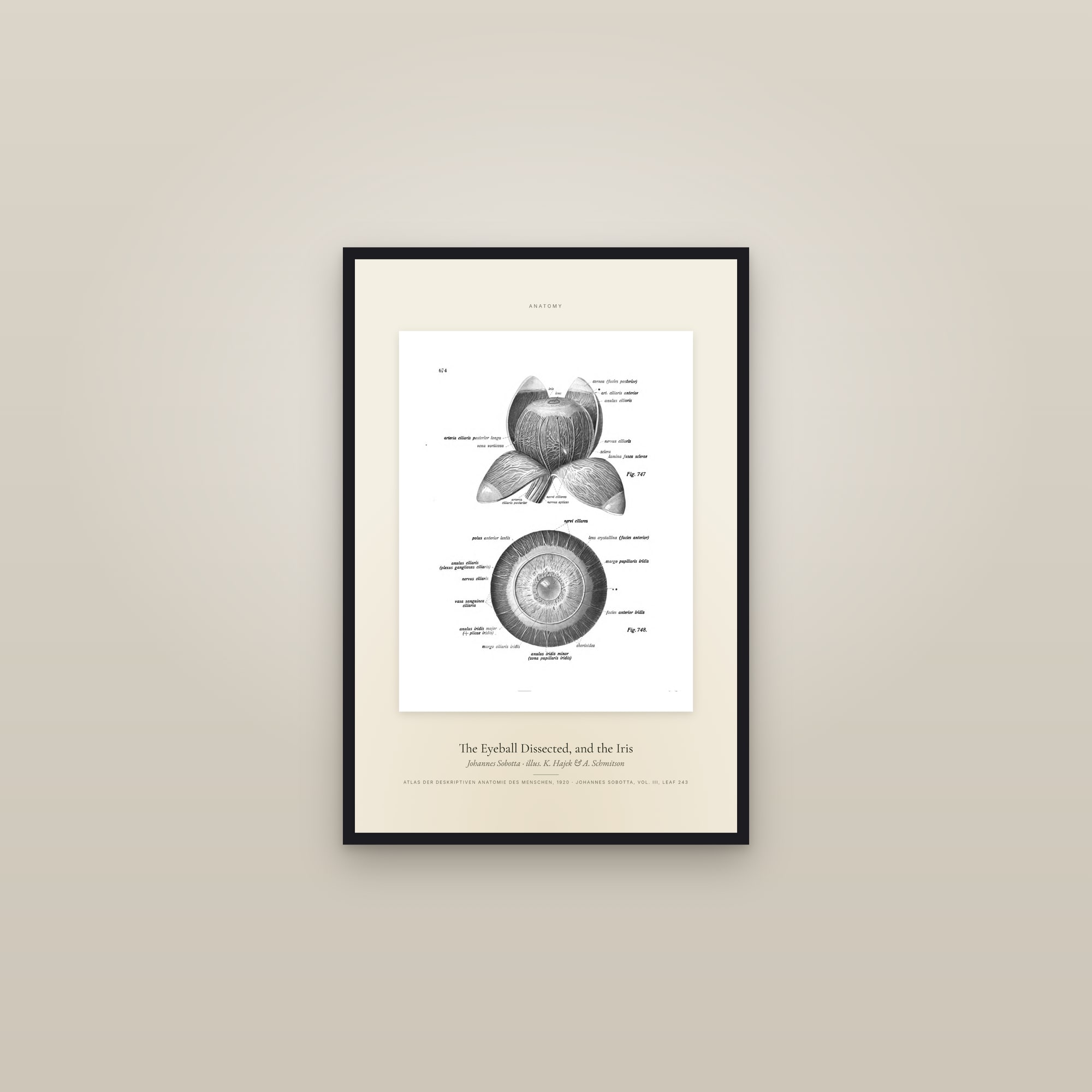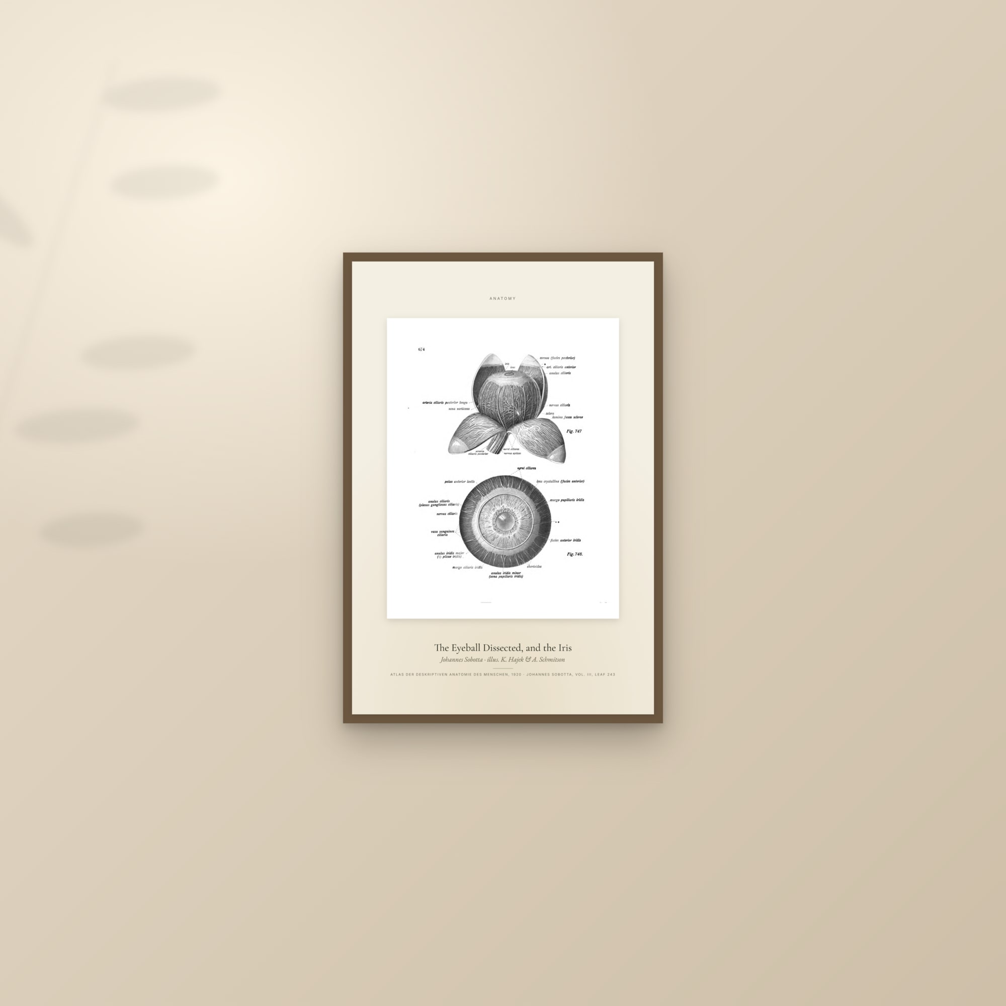
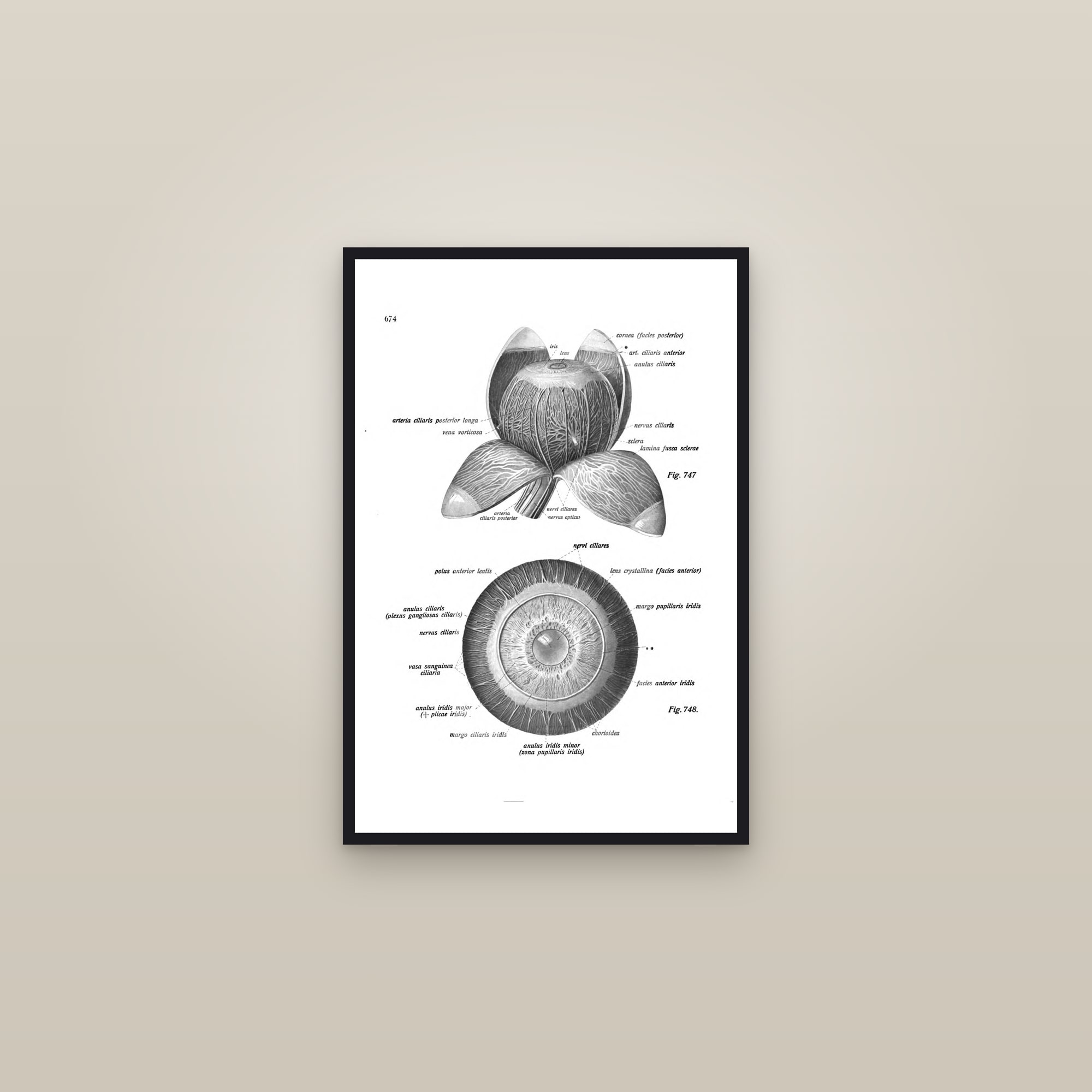
The Eyeball Dissected, and the Iris
The Eyeball Dissected, and the Iris (Johannes Sobotta · illus. K. Hajek & A. Schmitson) — Atlas der deskriptiven Anatomie des Menschen, 1920 · Johannes Sobotta, vol. III, leaf 243.
📦 Free shipping on every order, arriving in 4–9 business days with production included. Express ($14.90) at checkout arrives in 4–6 business days.
 This print
Where it works
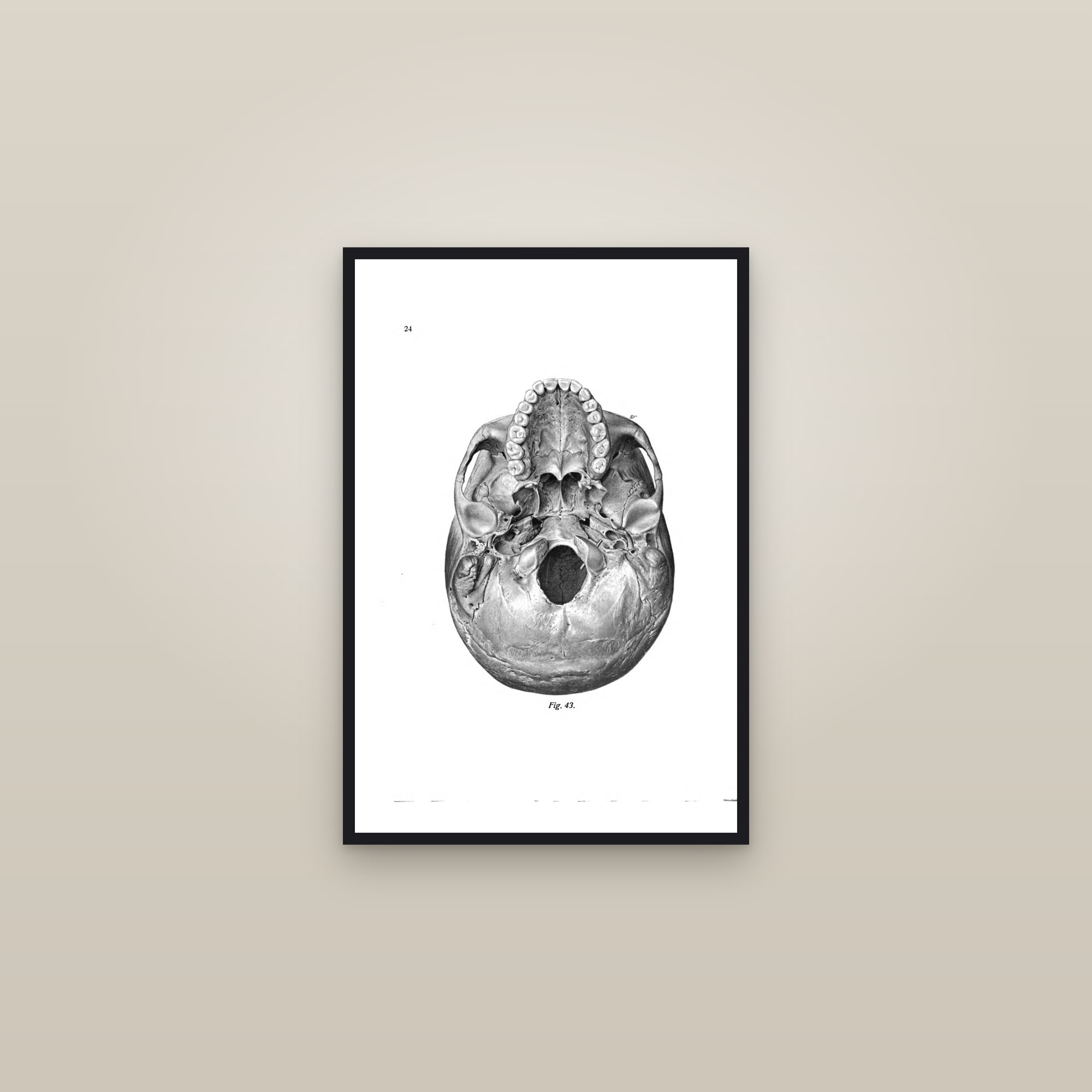
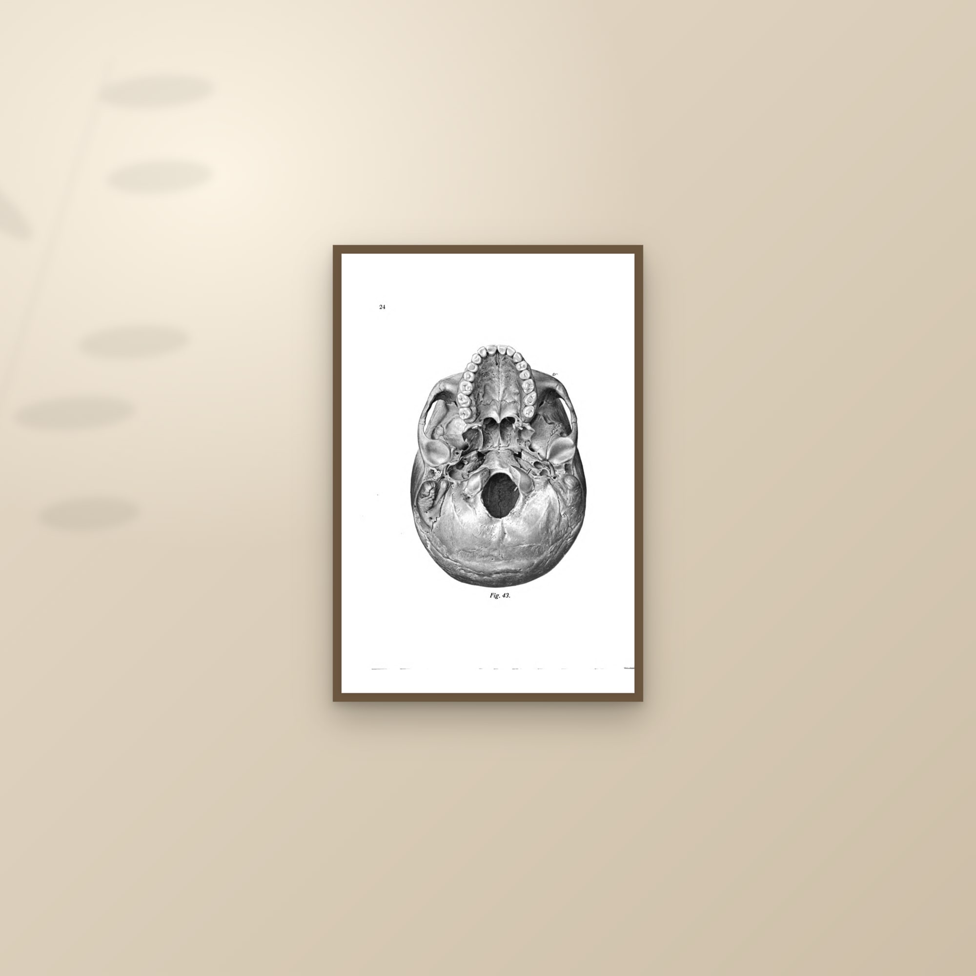
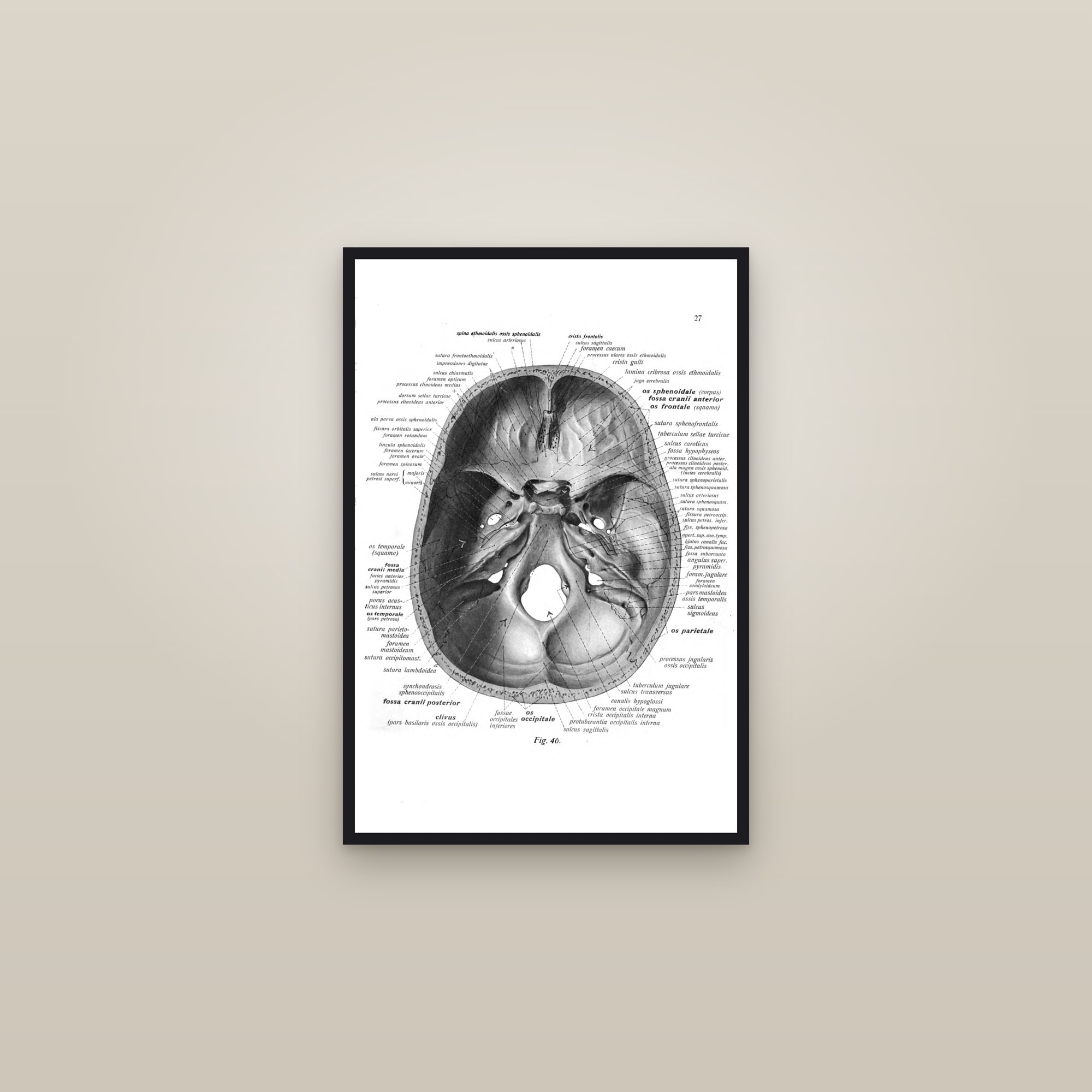
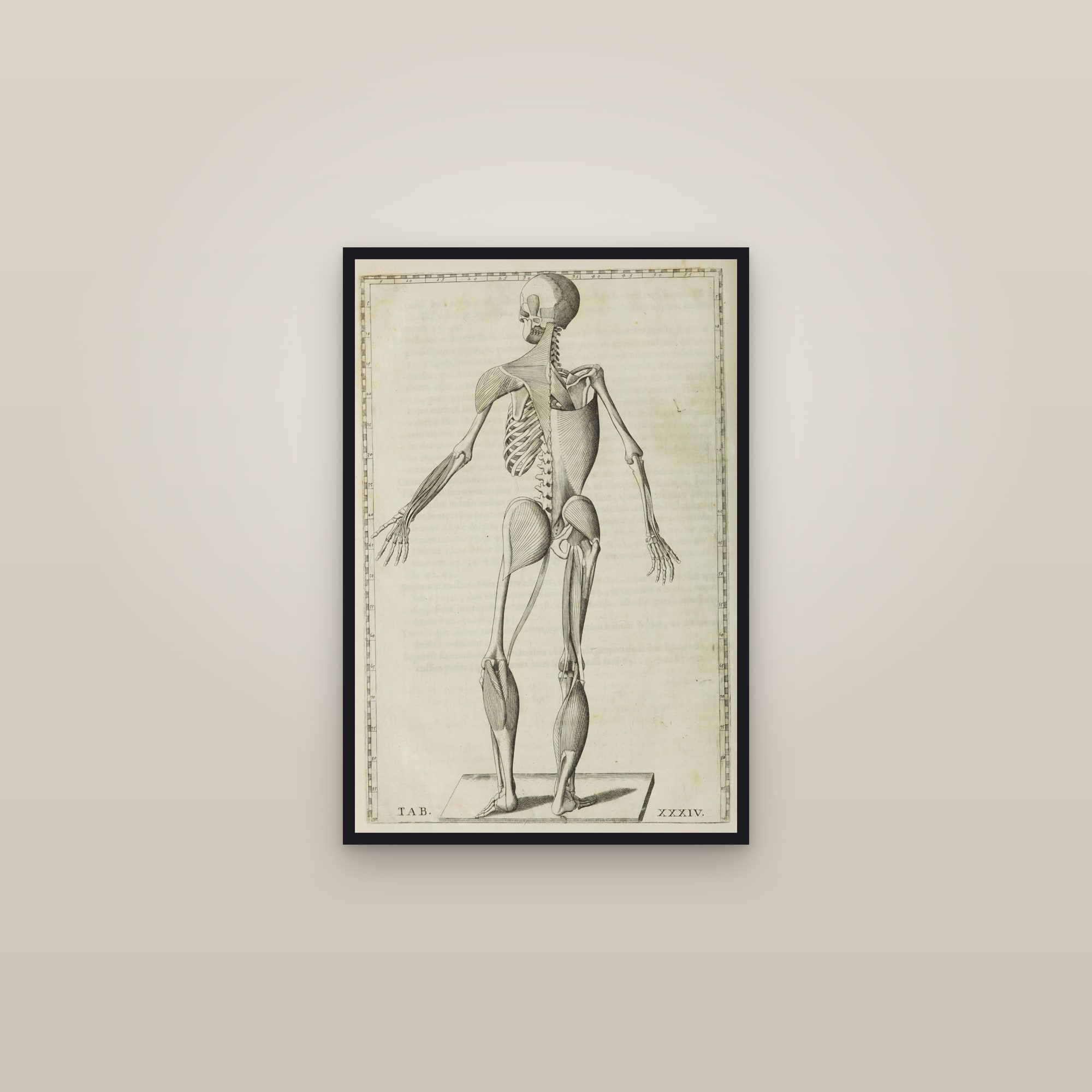
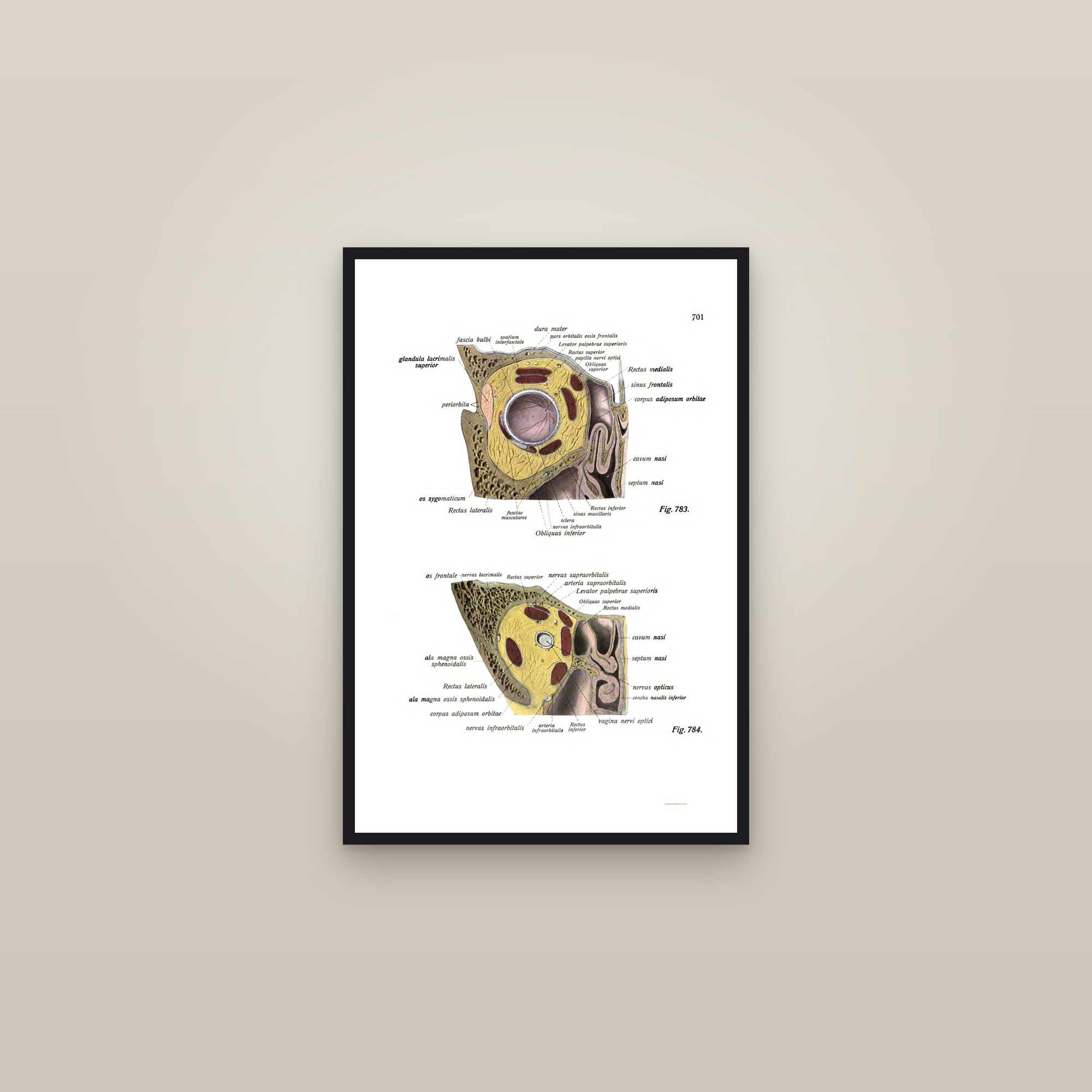
Sobotta's 1920 plate opens the eyeball like a flower, the white of the eye folded back to show the choroid vessels, the iris face-on below, in grey halftone. For an ophthalmologist's office, paired with The Eye in the Orbit (muscles of the eye), at 16×24 in a dark wood frame.
Style, size & framing detail
- Style
- Antique anatomical plate
- Era
- 19th century
- Orientation
- Portrait
- Palette
- grey, white
- Mood
- calm, scholarly
- Rooms
- home office, study or library, classroom
- Good gift for
- scientists, men, him, doctors & nurses
Size. We'd hang this one at 16×24. Prints at 12×18, 16×24, 24×36 in — 12×18 for a shelf, desk or a gallery-wall grid; 16×24 above a desk, console or in a hallway; 24×36 as the statement piece above a sofa or bed.
Framing. A slim black or dark wood frame with no mat (black sharpens an engraving, dark wood warms a colour plate), hung at eye level so the plate can be read. Also available gallery-wrapped on canvas, ready to hang with no frame.
Pairing. Hangs alone or as a matched pair; three or more prints are 10% off.
Details & materials
Shipping & returns
About this design
The Madten promise
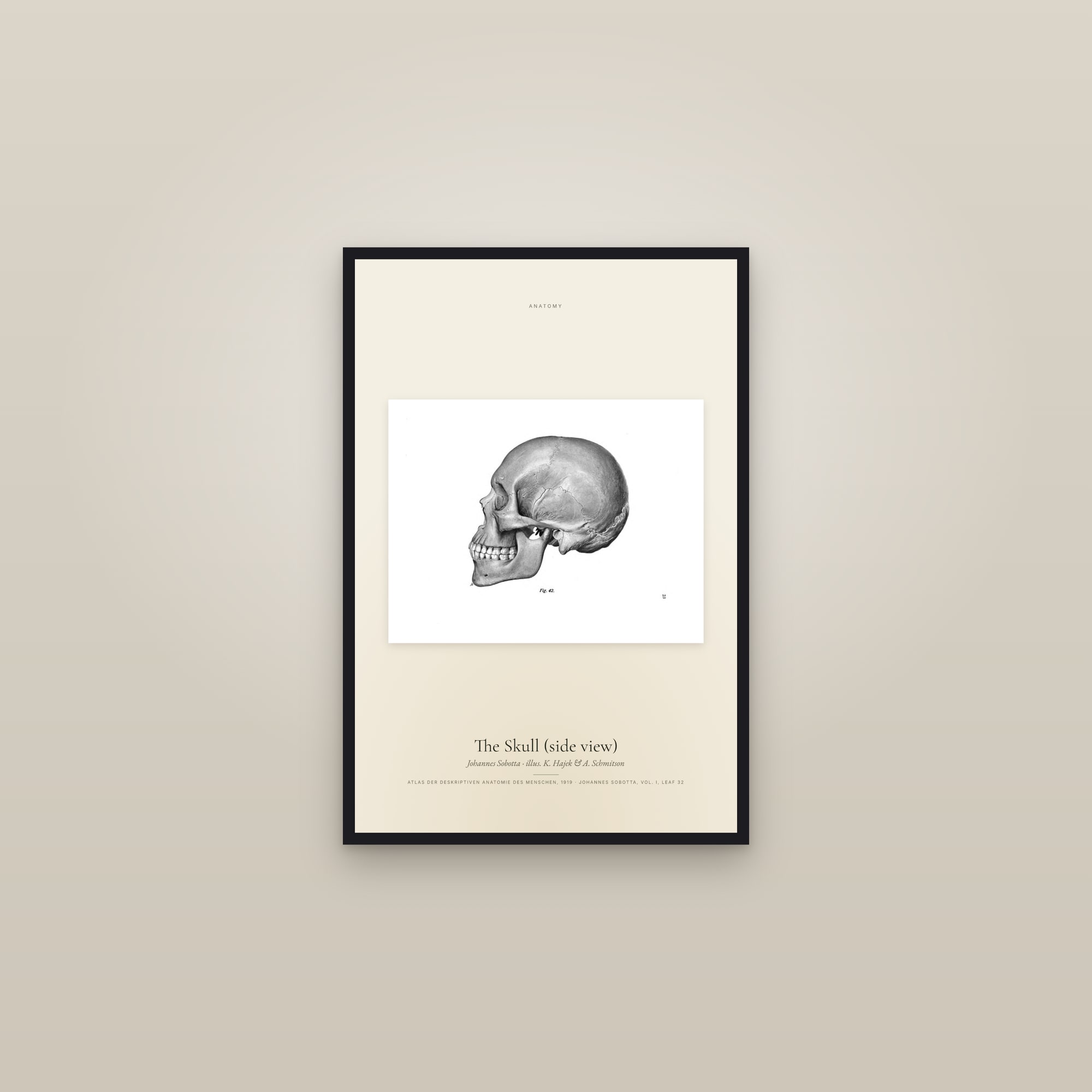
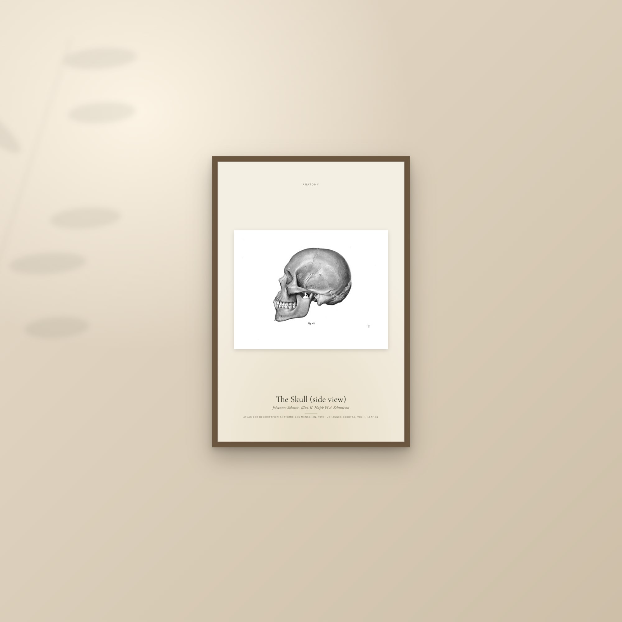
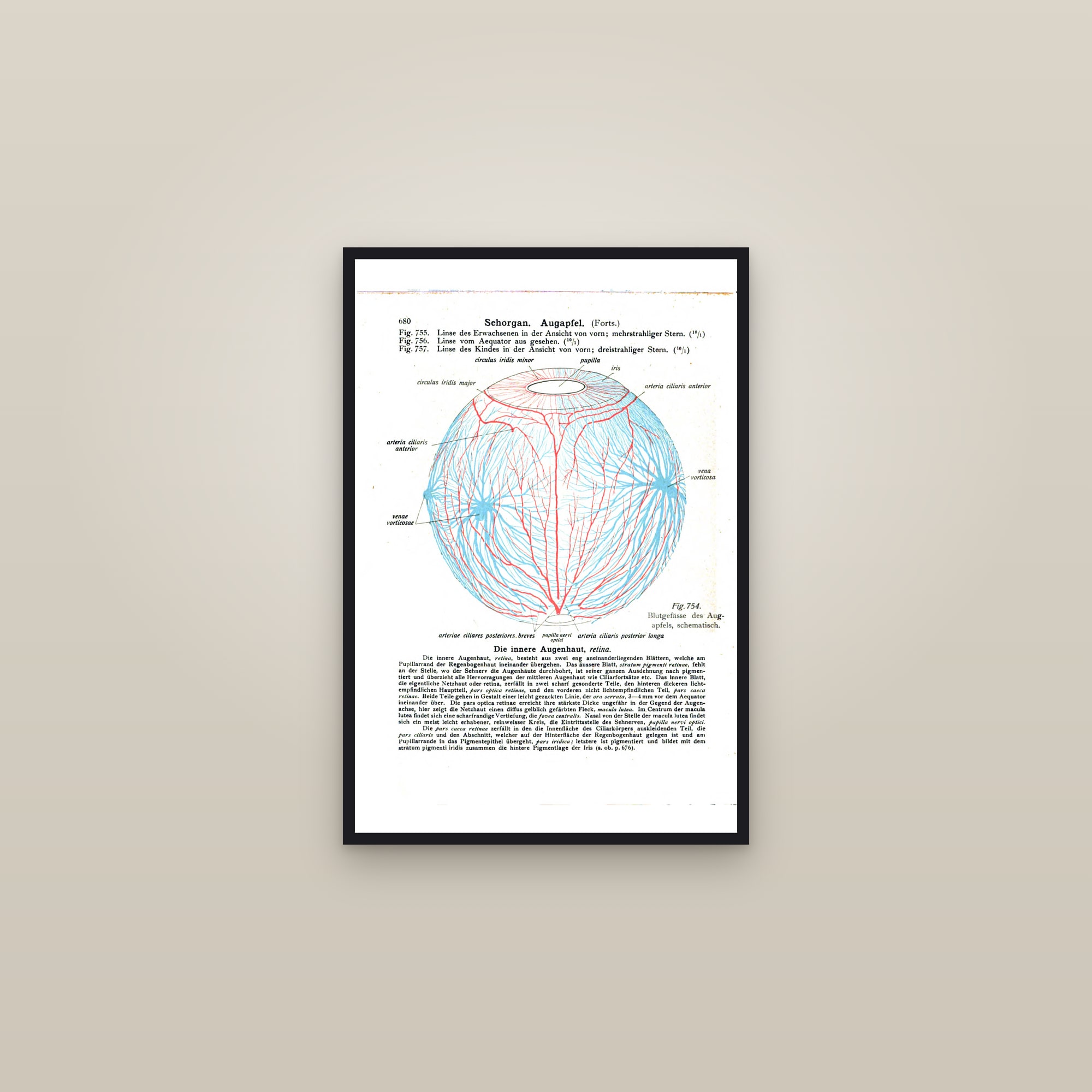
The cream specimen edition
The same plate set on a soft cream ground with a typeset specimen label — a gallery-ready print.