
Historical anatomy plates, 1543–1920, by body part
151 plates from five anatomical atlases, from Vesalius’s woodcuts of 1543 to Sobotta’s colour atlas of 1920. Each is arranged by the part of the body it shows, with its source, its place in the book and its date.
For four centuries the anatomical atlas was where medicine and art met. Anatomists dissected and described; artists, engravers and printers turned what they saw into pictures precise enough to teach from and beautiful enough to collect. This page gathers the plates we have restored from five of those atlases and arranges them by body part, so the same structure can be compared across 377 years: the heart as Eustachi engraved it in 1552 beside the heart as Sobotta’s artists painted it in 1920.
Every plate is in the public domain. Each entry gives the atlas, the plate’s place in the book as the original records it, and the year, so it can be found in the digitised originals listed under each atlas below. The pictures on this page are free to view.
The five atlases
In the order they were made. Each lists who made it and where to see the original.
- 1543
De humani corporis fabrica
Andreas Vesalius (1514–1564)
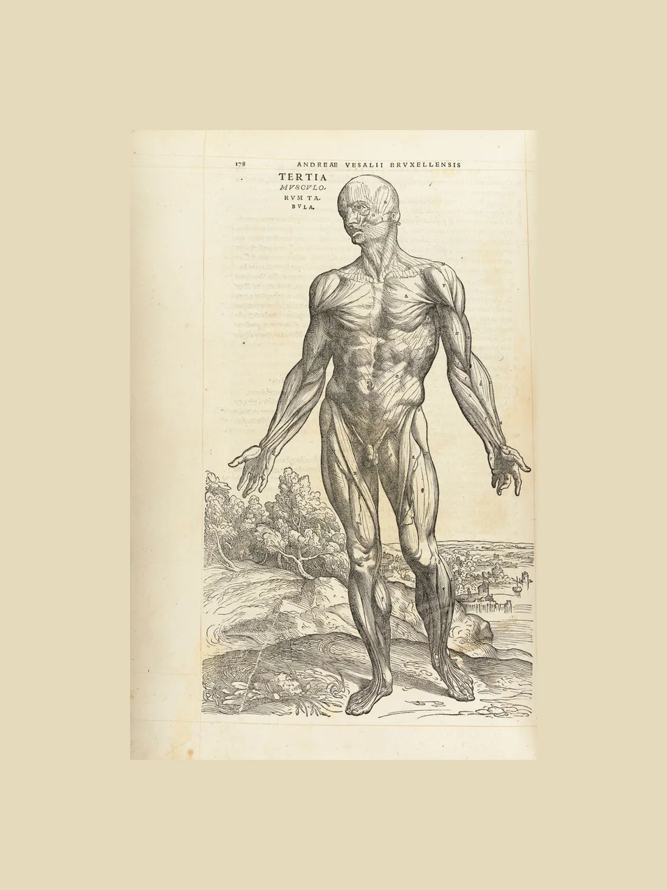
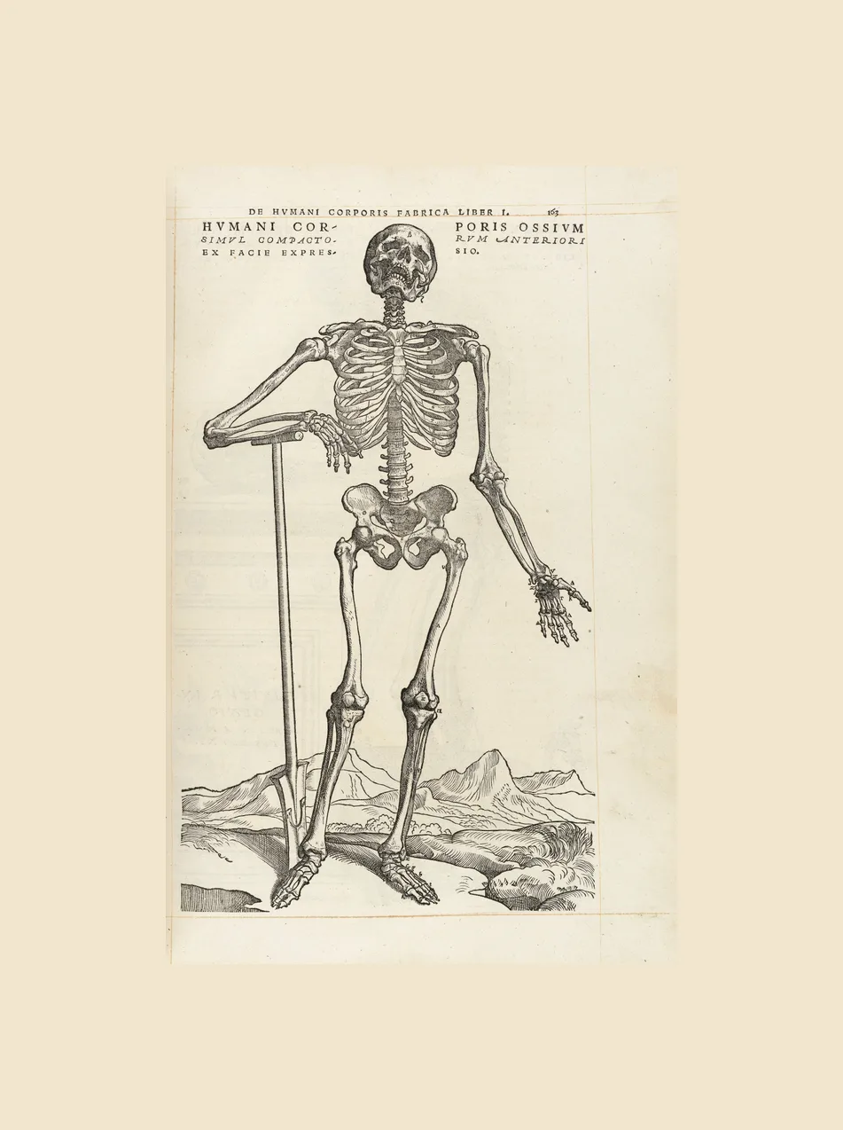
Vesalius was 28 and professor of anatomy at Padua when the Fabrica appeared. He built it on his own dissections rather than on the ancient authority of Galen, and the book is usually taken as the beginning of modern anatomy.
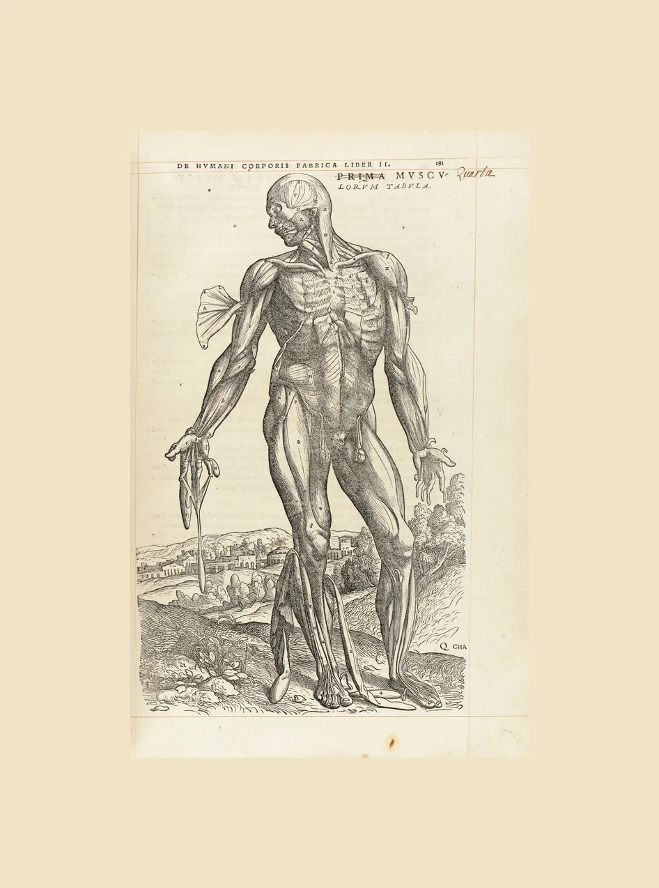
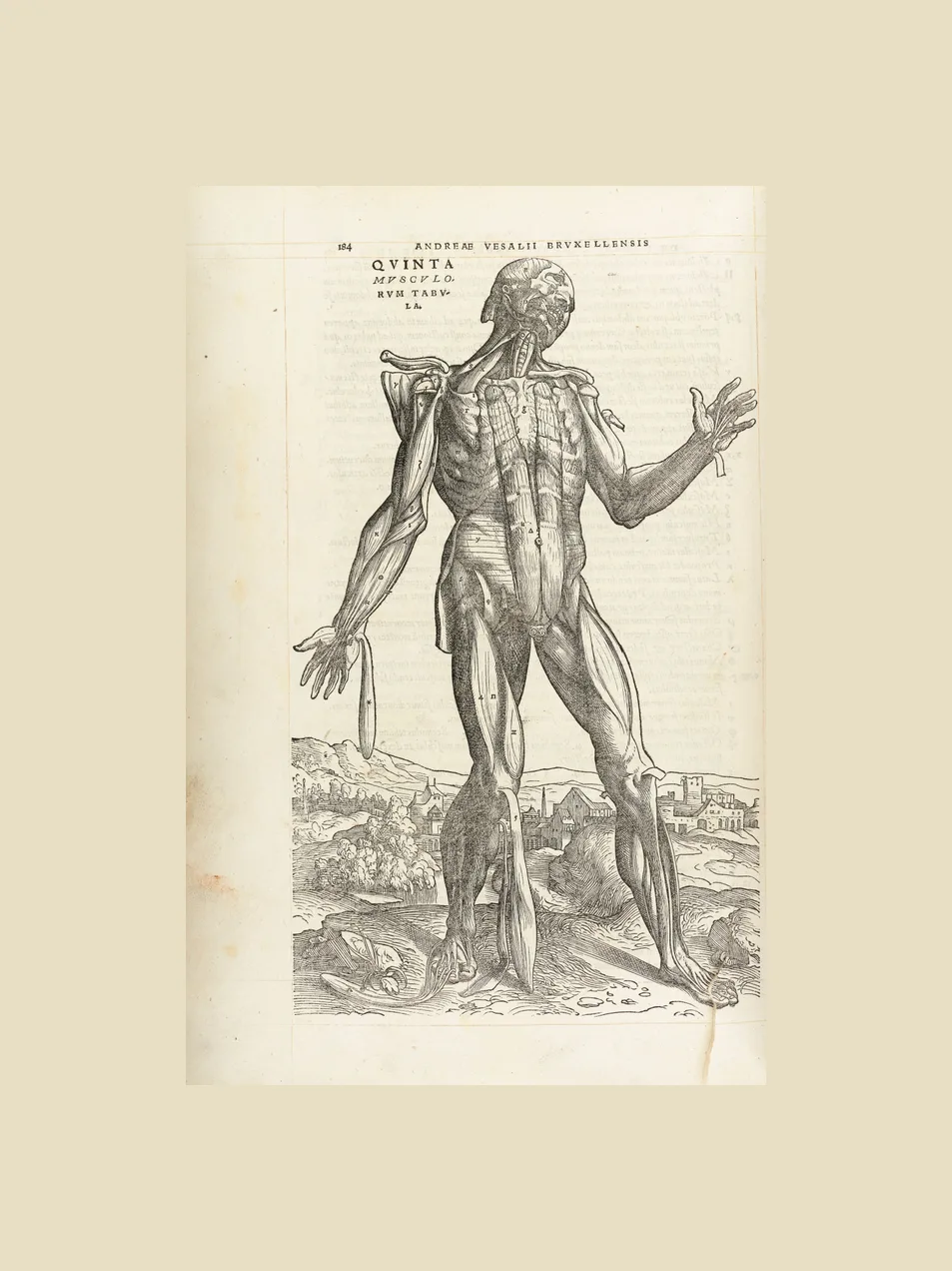
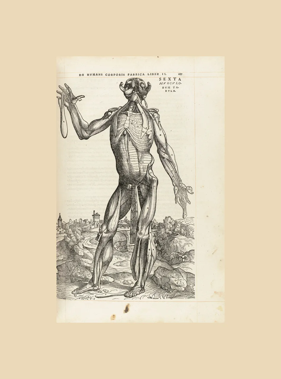
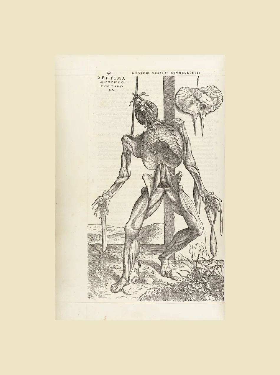
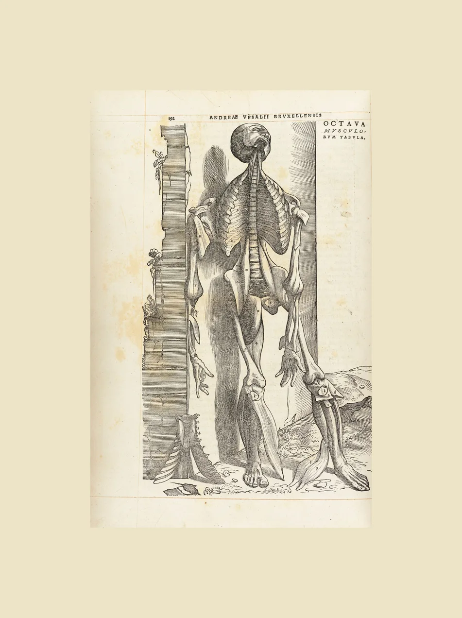
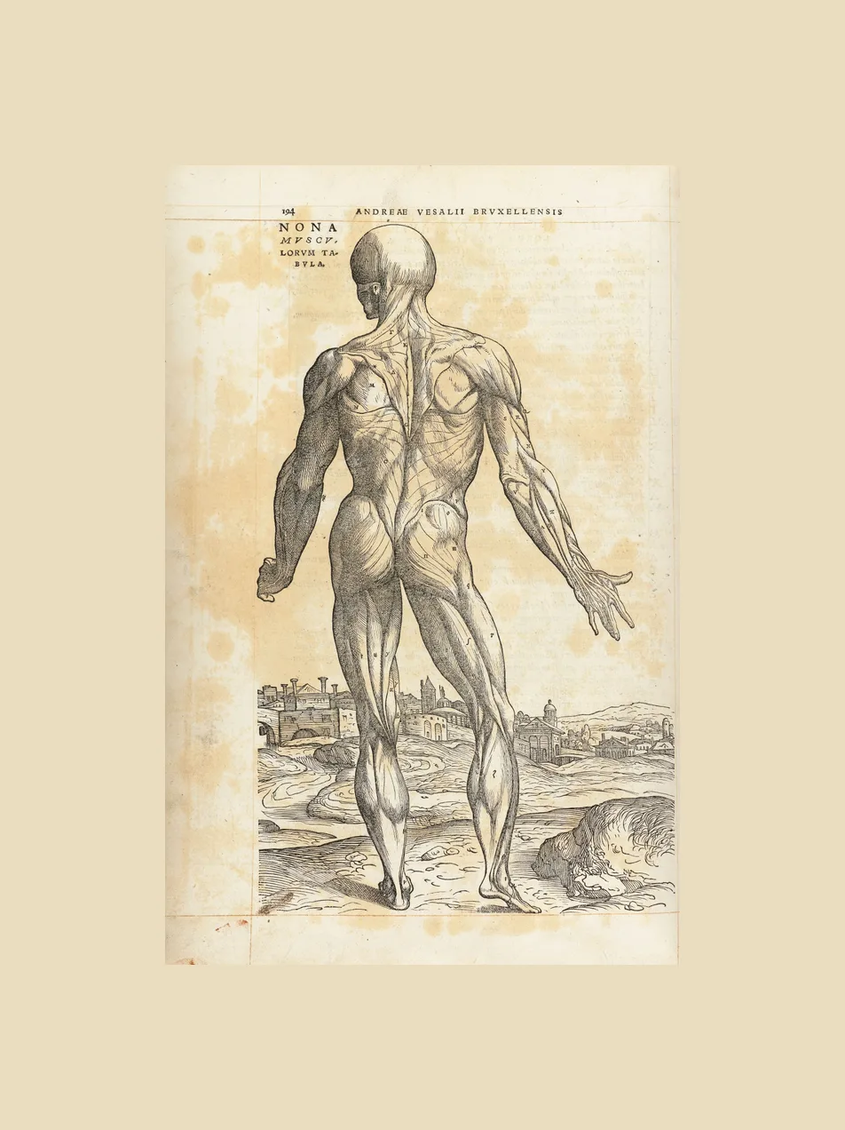
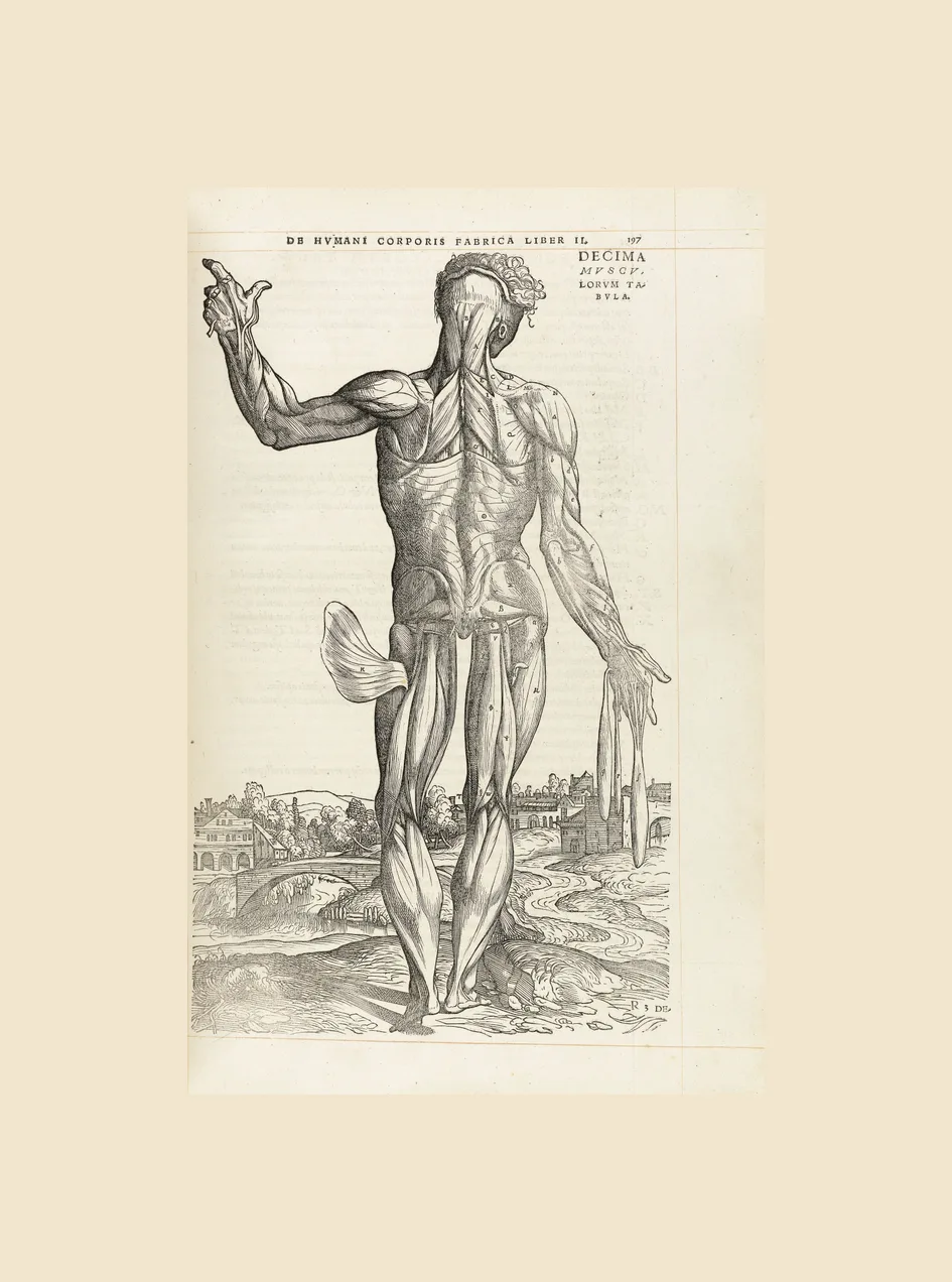
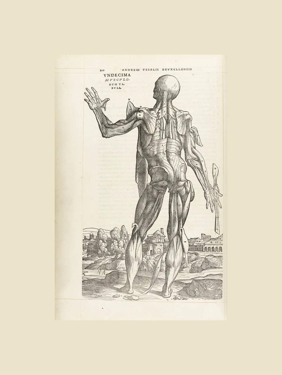
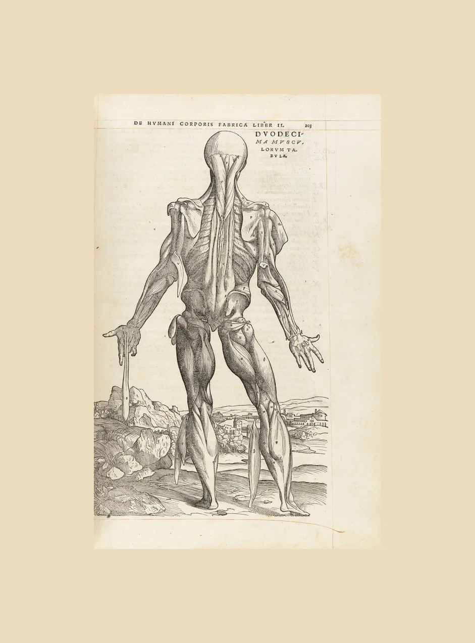
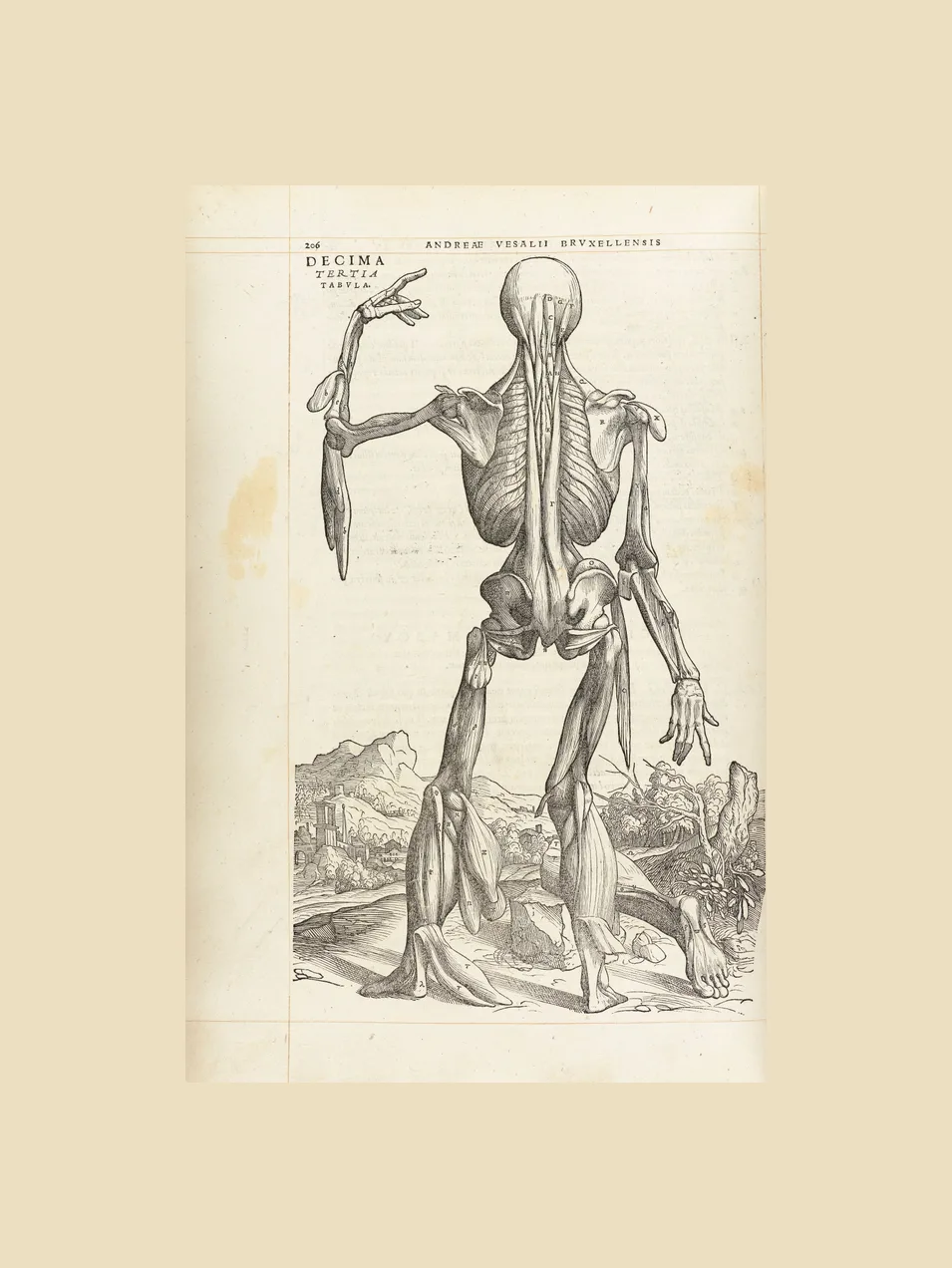
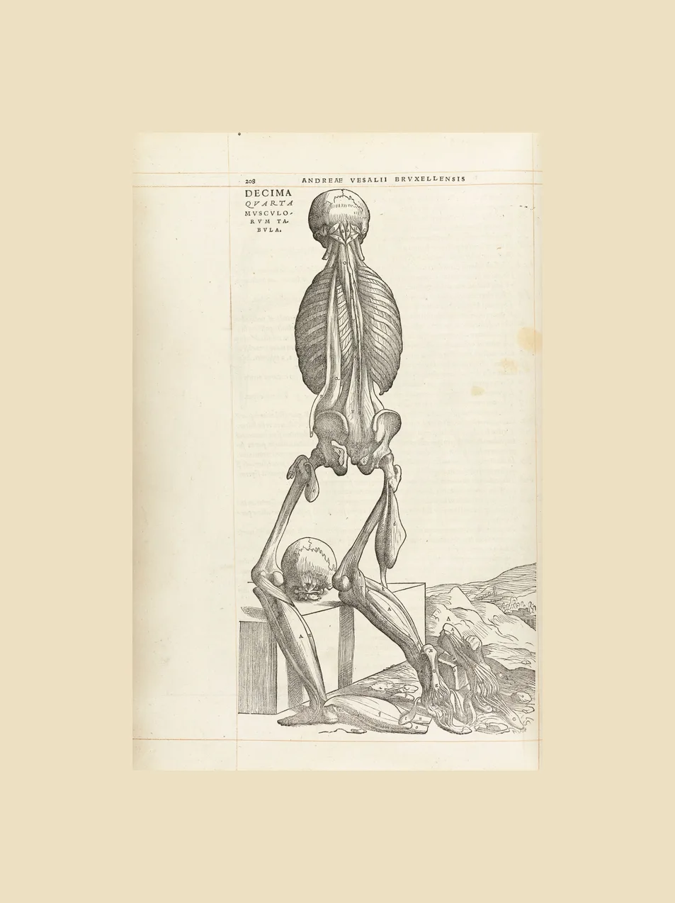
Its best-known woodcuts are the fourteen “muscle men”, figures that peel away layer by layer while posing in a landscape. Set side by side, the backgrounds of several of them join into one continuous view of the hills near Padua. The original woodblocks survived until 1944, when they were destroyed in the bombing of Munich.
- Made by
- Woodcuts from the workshop of Titian, often attributed to Jan Steven van Calcar
- Published
- Basel: Johannes Oporinus, 1543
- Medium
- Woodcut
- On this page
- 21 plates
- See the original
- National Library of Medicine, Historical Anatomies on the Web
- 1552 / 1714
Tabulae anatomicae
Bartolomeo Eustachi (died 1574)
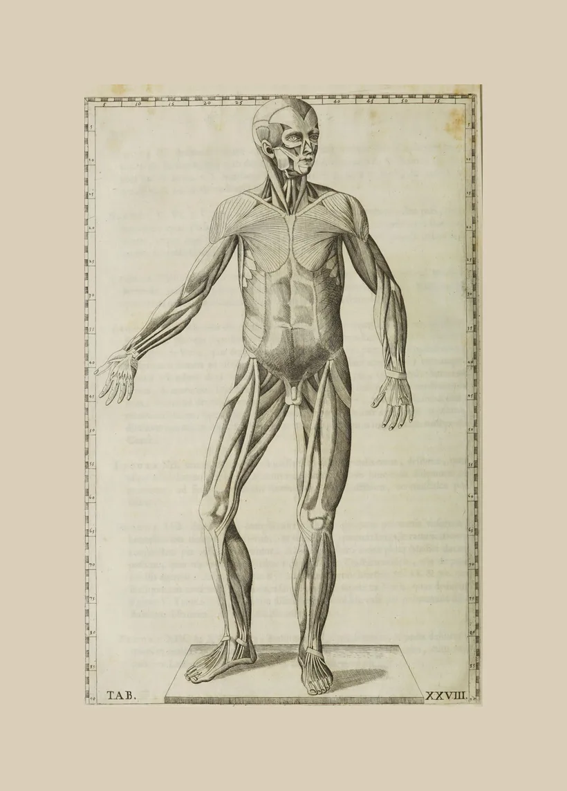
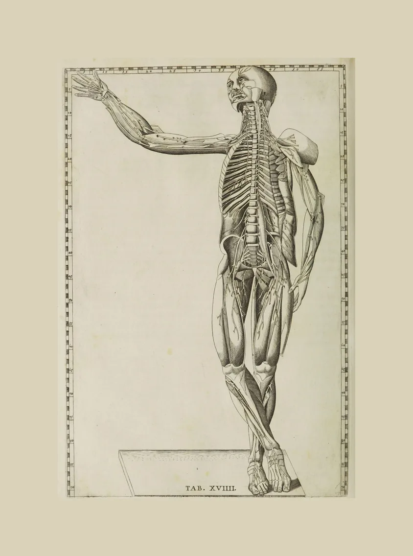
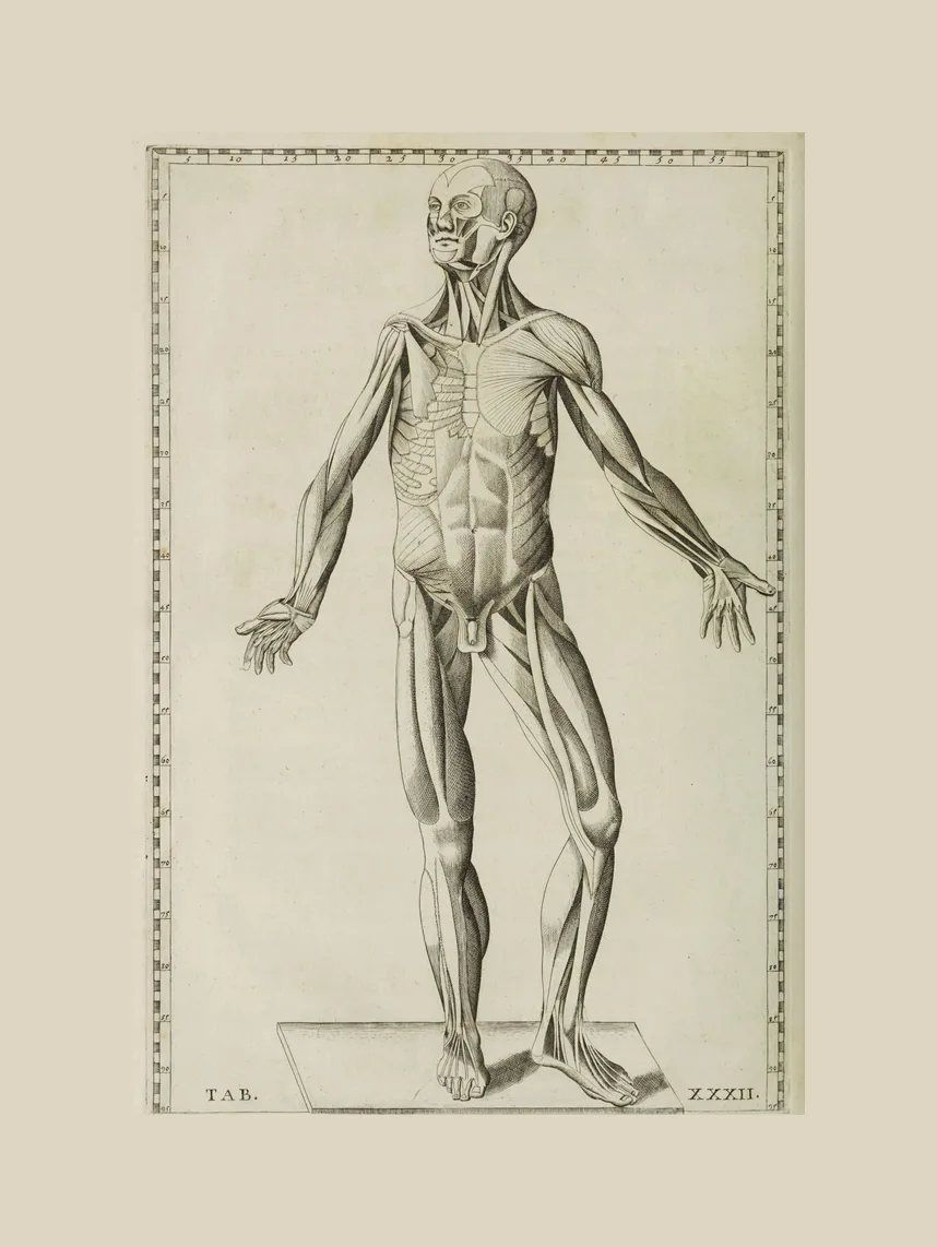
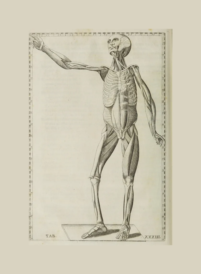
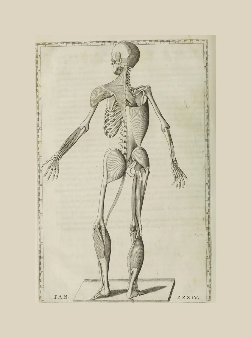
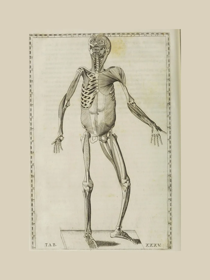
Eustachi, the anatomist who gave his name to the Eustachian tube, had forty-seven copper plates engraved in Rome in 1552. Only eight were printed in his lifetime. The rest lay unpublished for more than 150 years, until the papal physician Giovanni Maria Lancisi brought them out as a complete set in 1714.
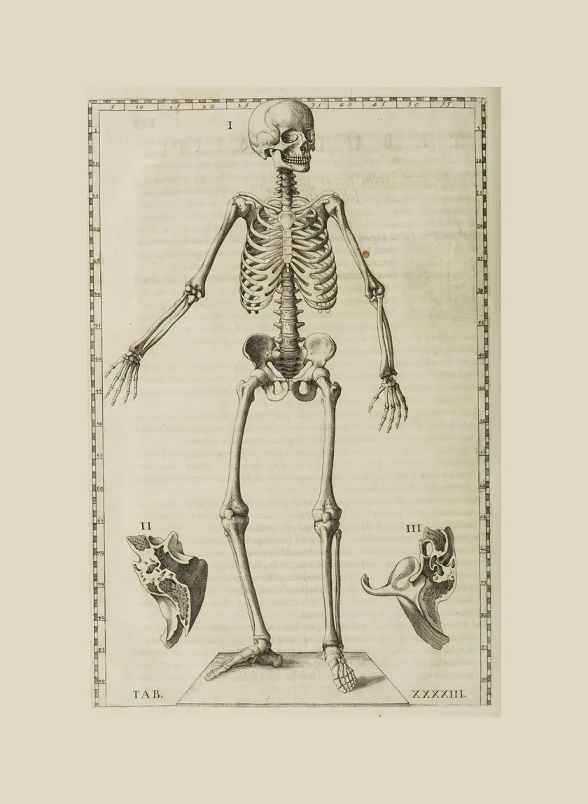
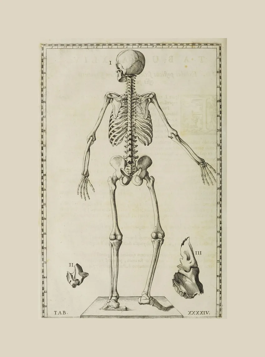
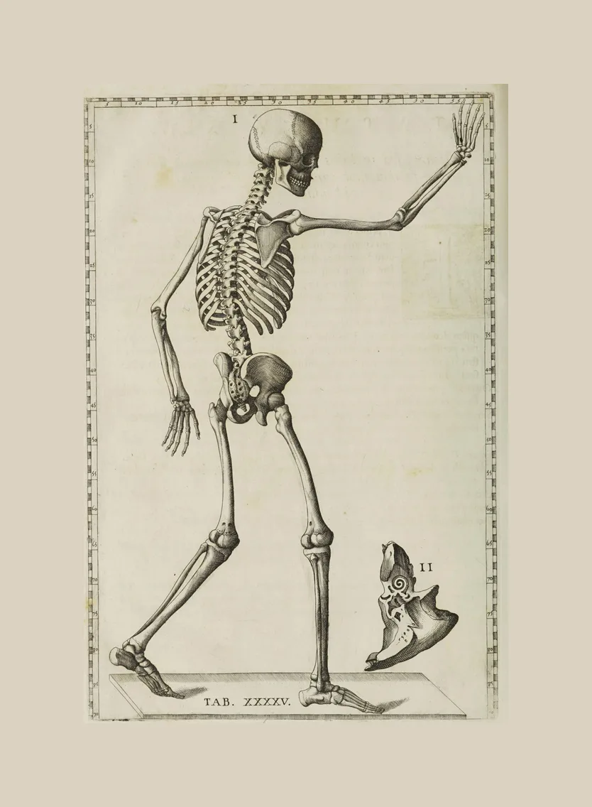
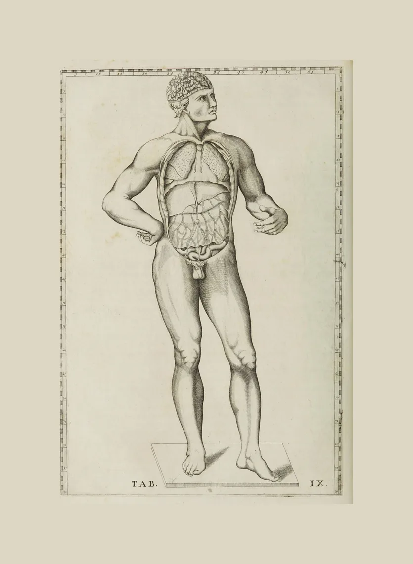
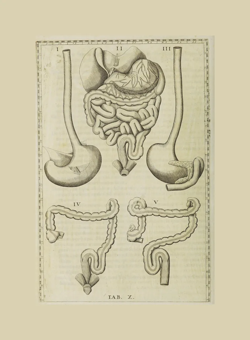
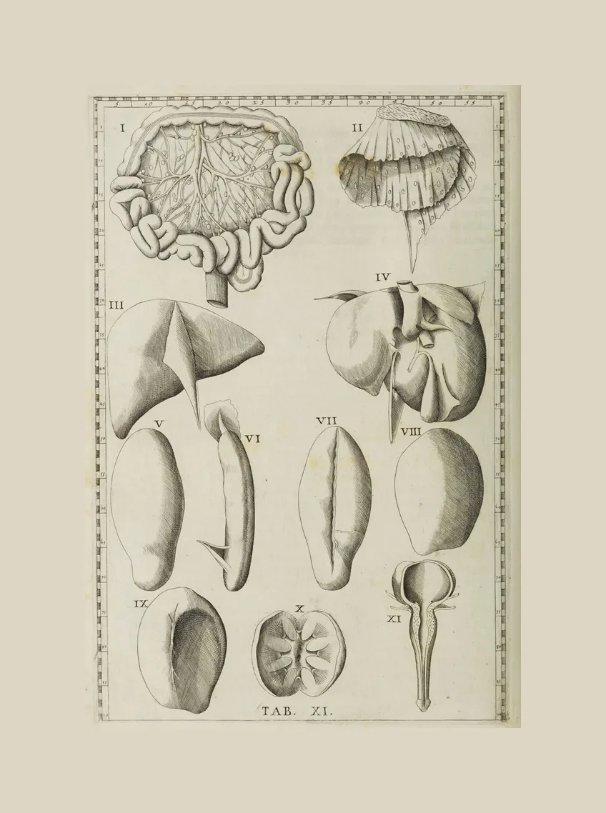
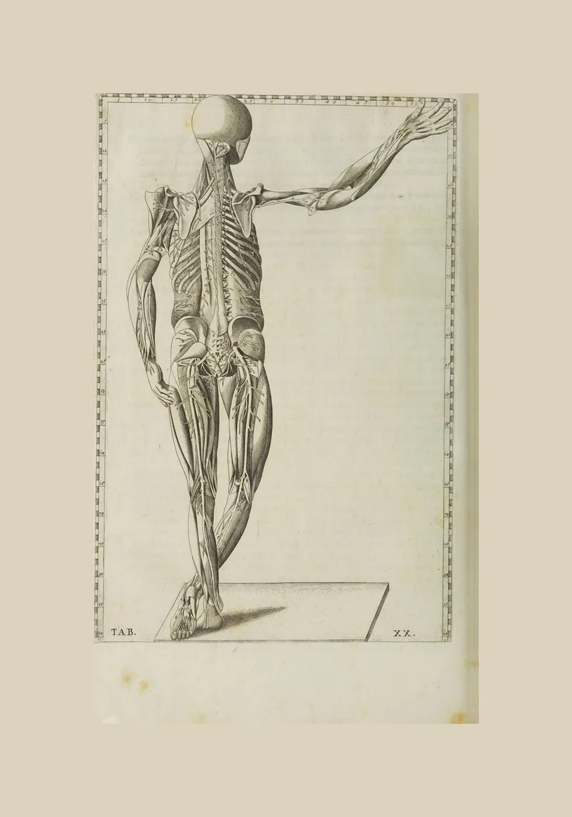
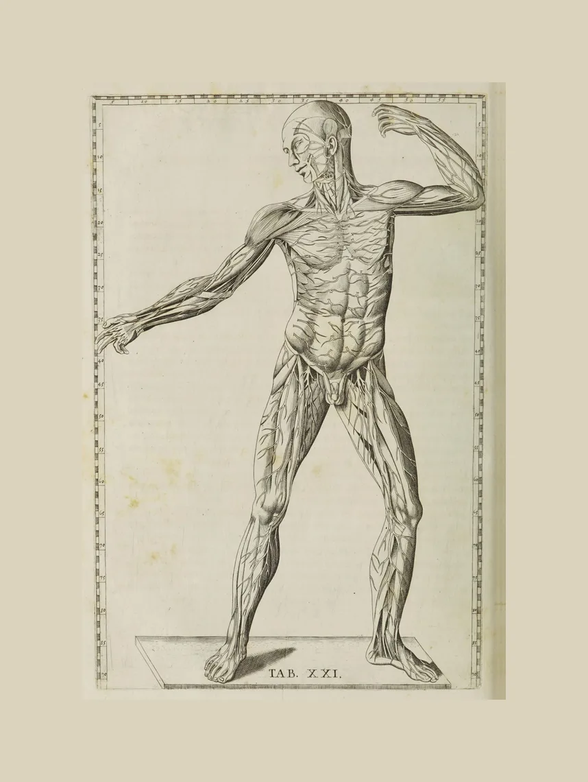
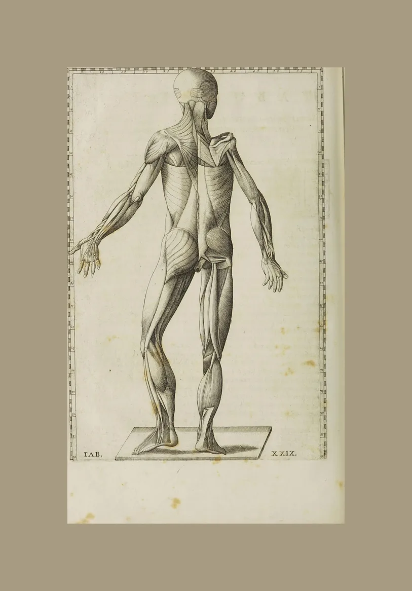
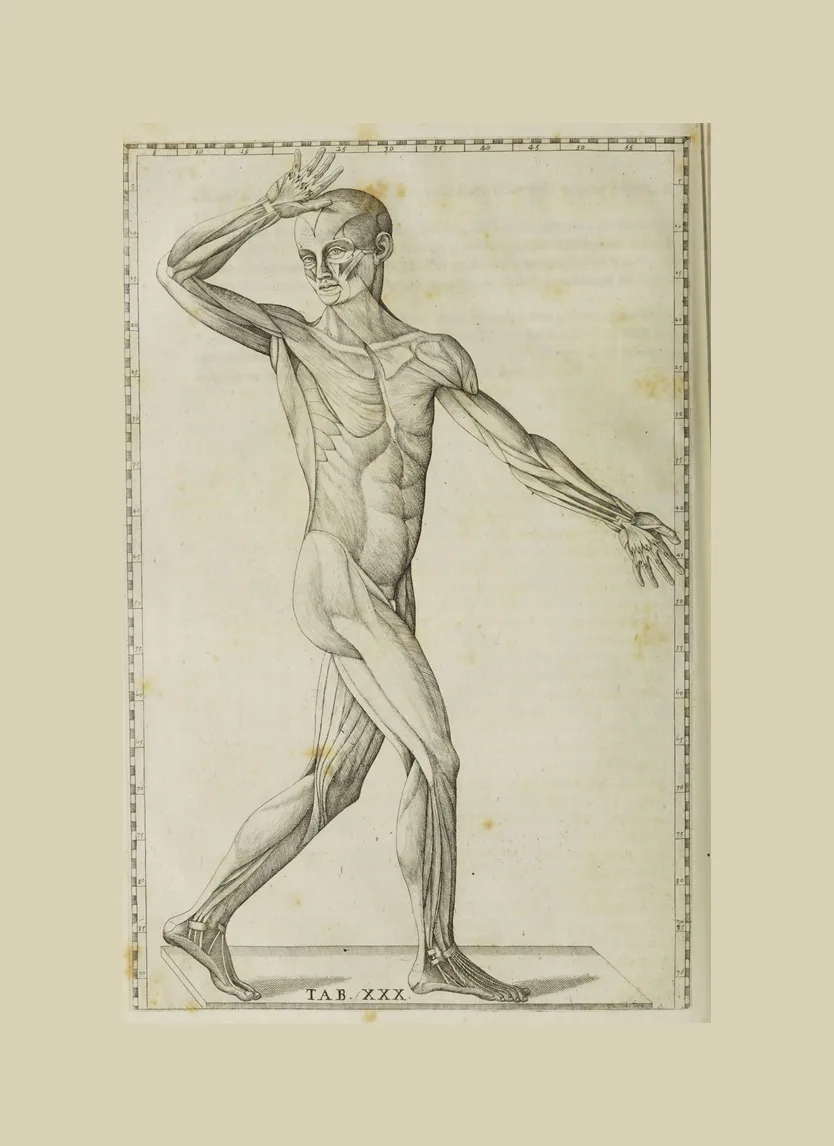
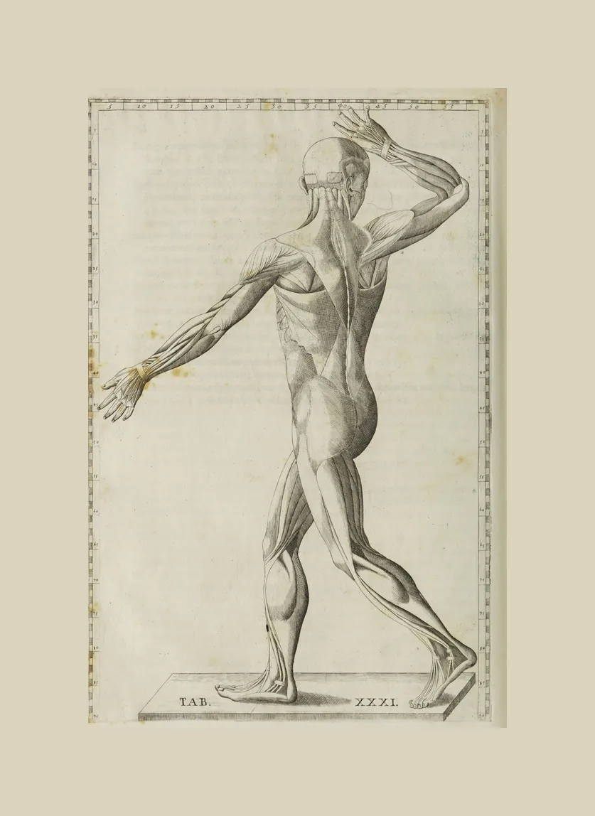
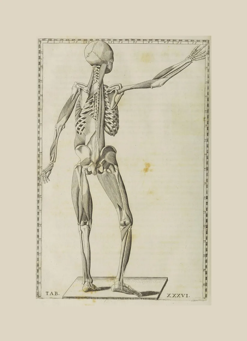
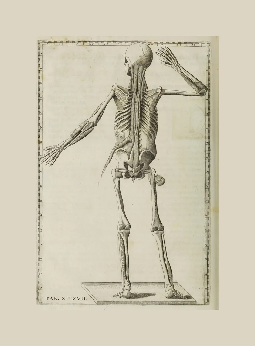
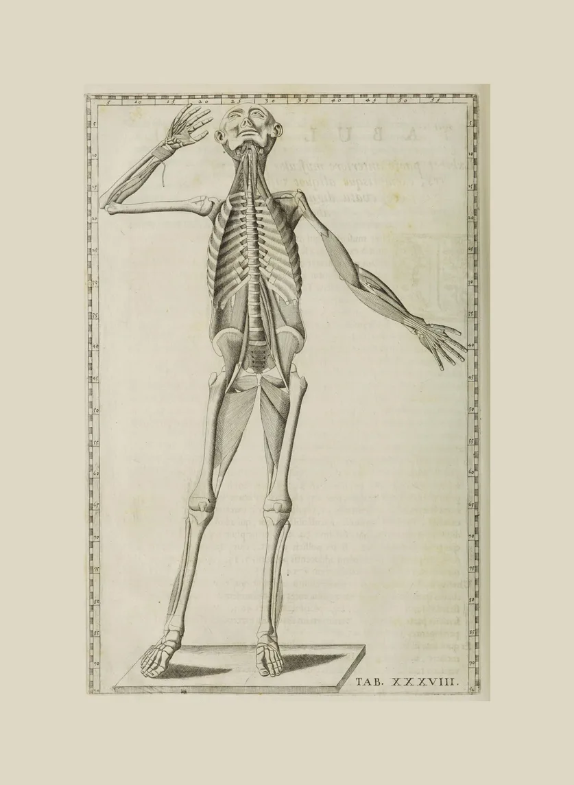
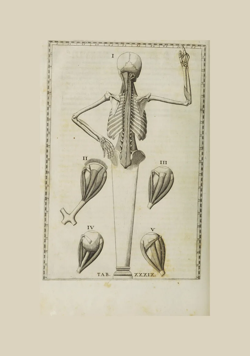
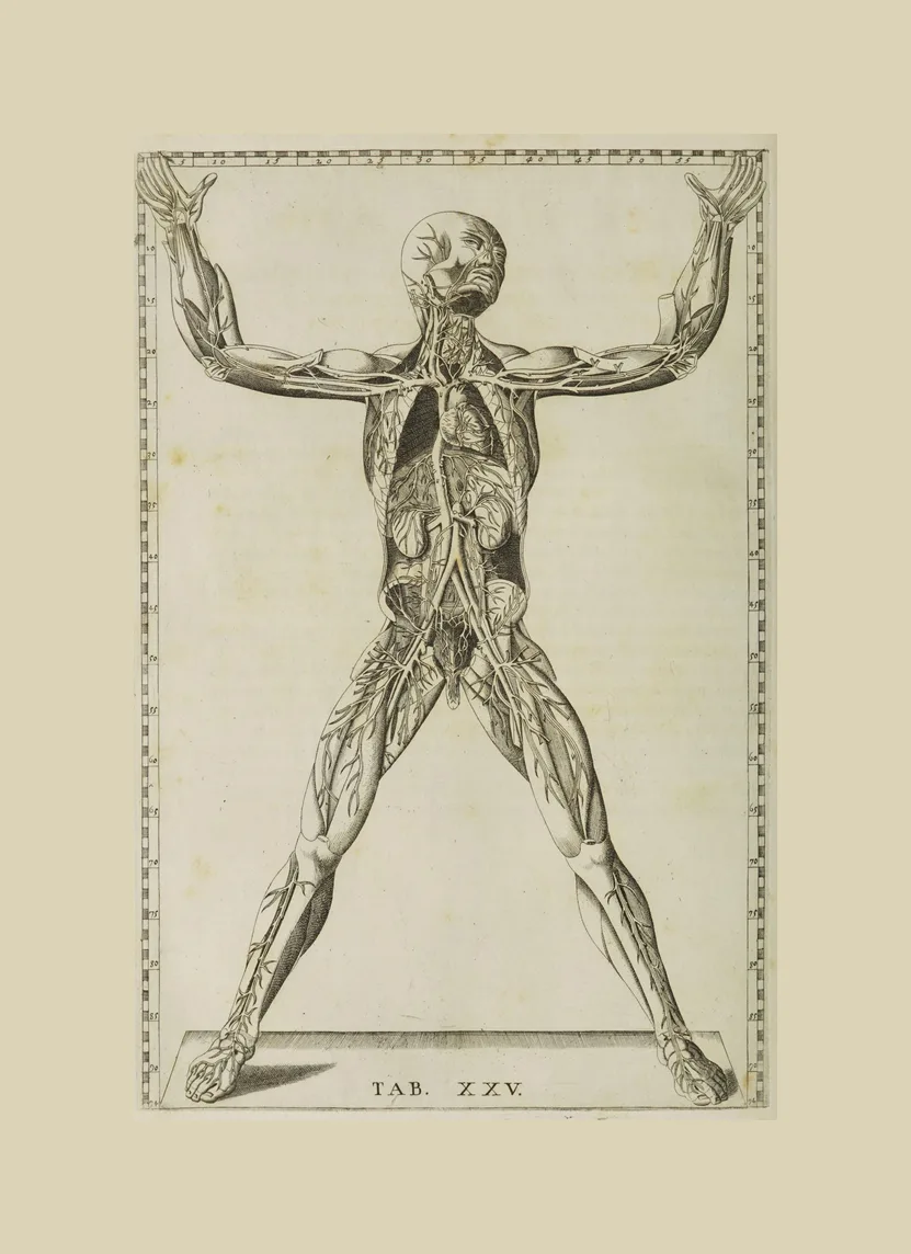
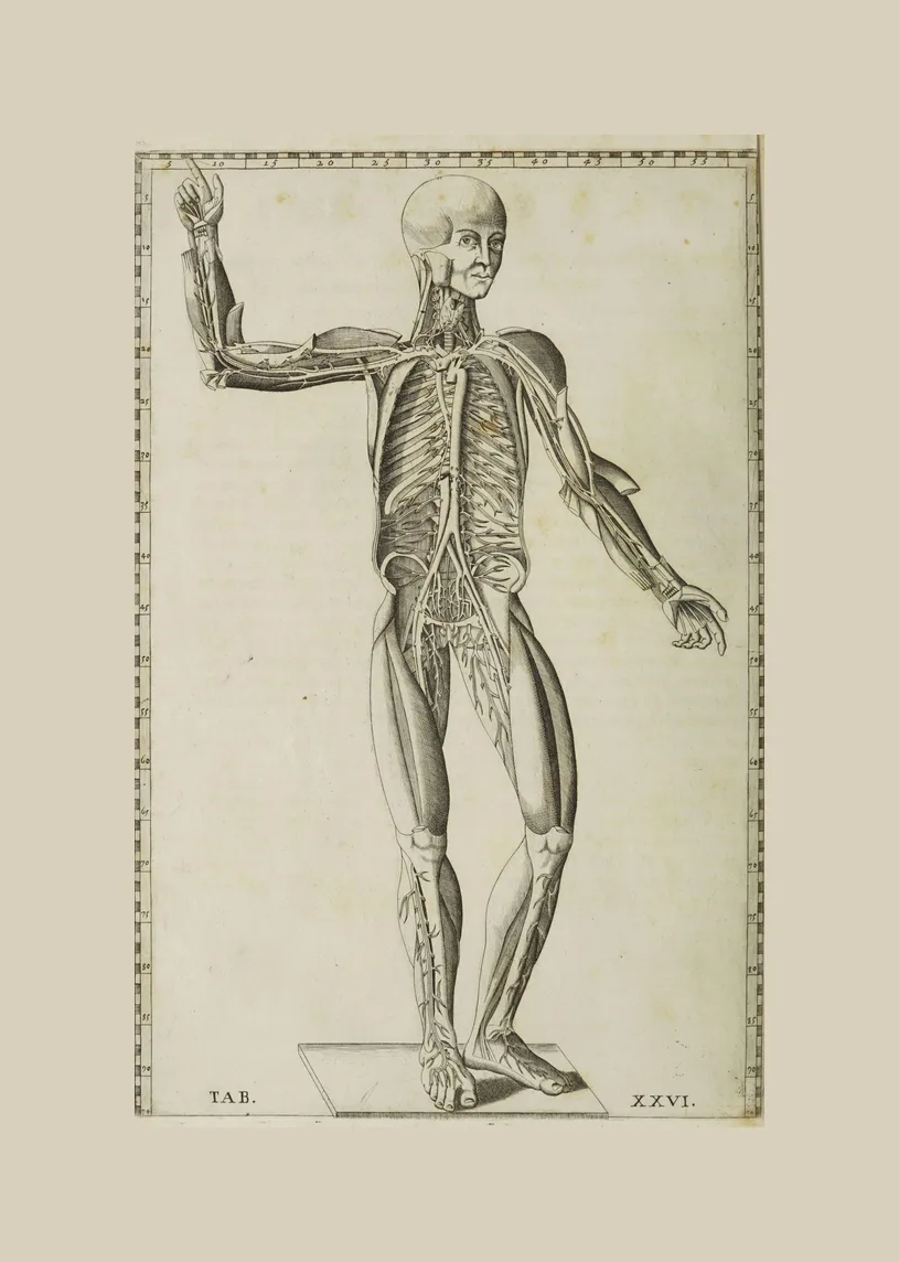
The figures carry no labels. Instead each one stands inside a ruled border marked with numbers, so a reader can locate any structure by its coordinates, like a map. That is why the plates still read so cleanly today.
- Made by
- Copper engravings by Giulio de’ Musi
- Published
- Rome: edited by Giovanni Maria Lancisi, engraved 1552, published 1714
- Medium
- Copper engraving
- On this page
- 39 plates
- See the original
- National Library of Medicine, Historical Anatomies on the Web
- 1747
Tabulae sceleti et musculorum corporis humani
Bernhard Siegfried Albinus (1697–1770)
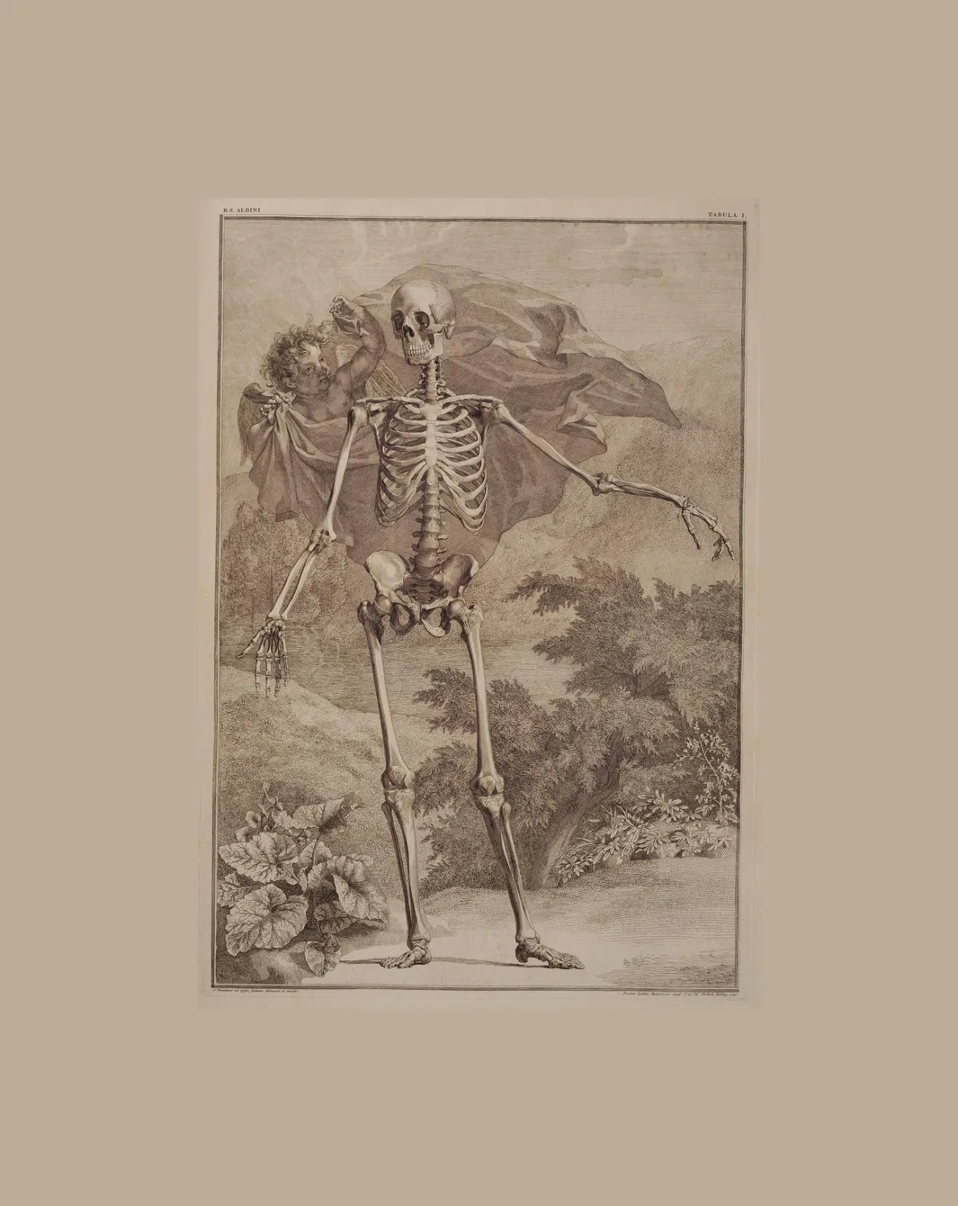
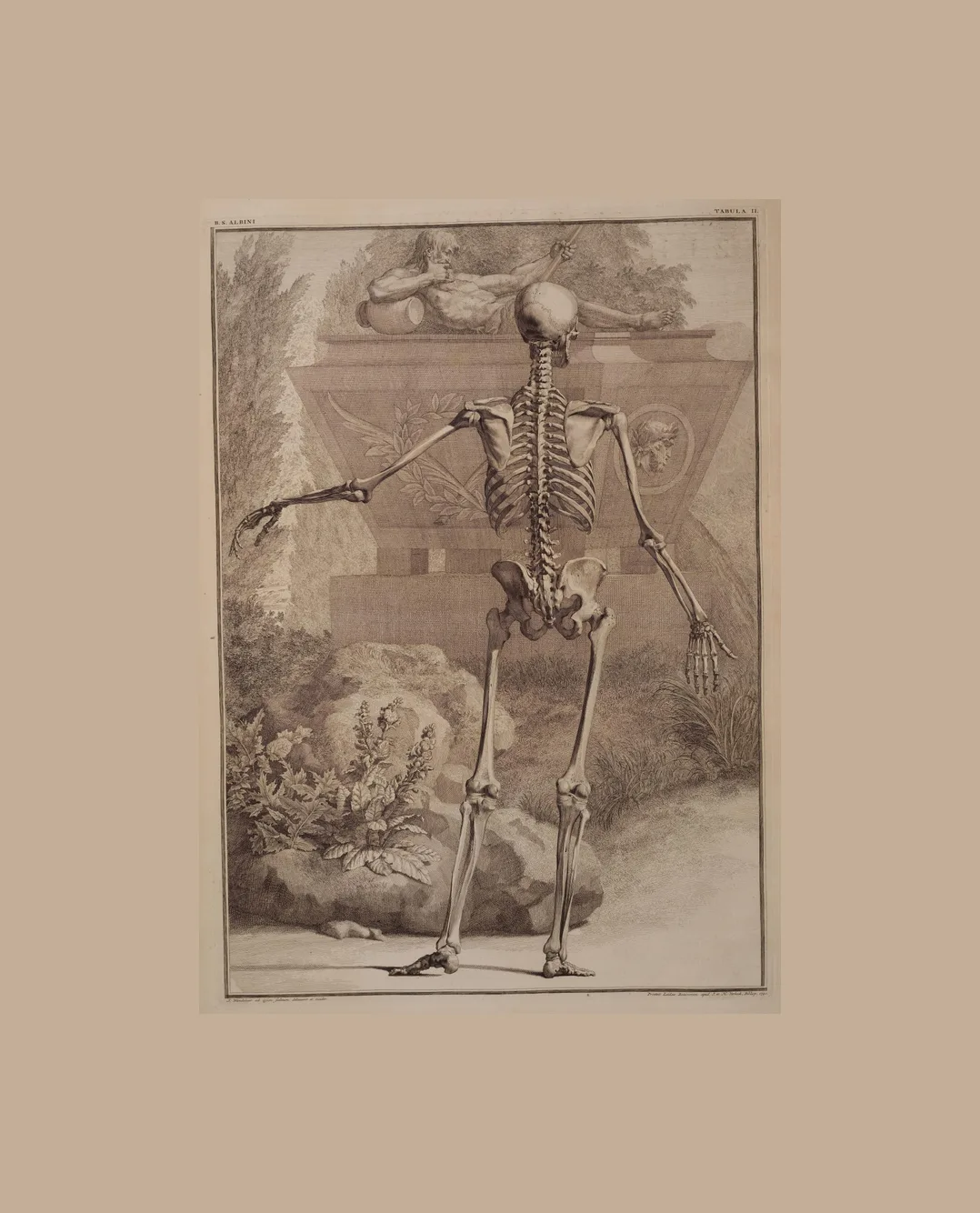
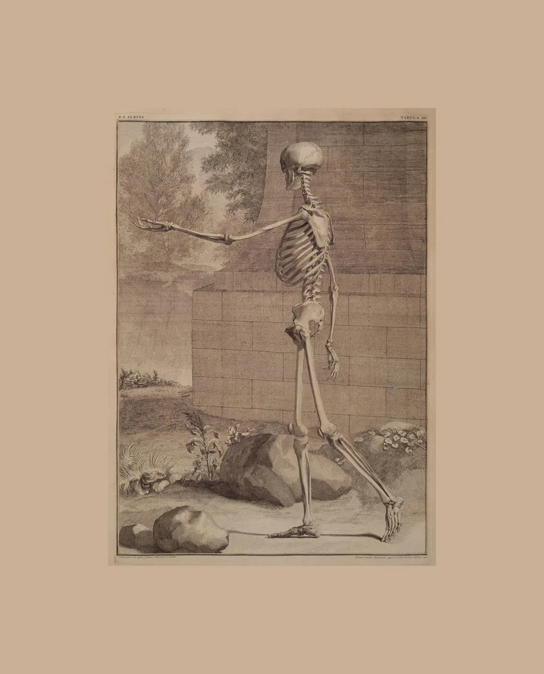
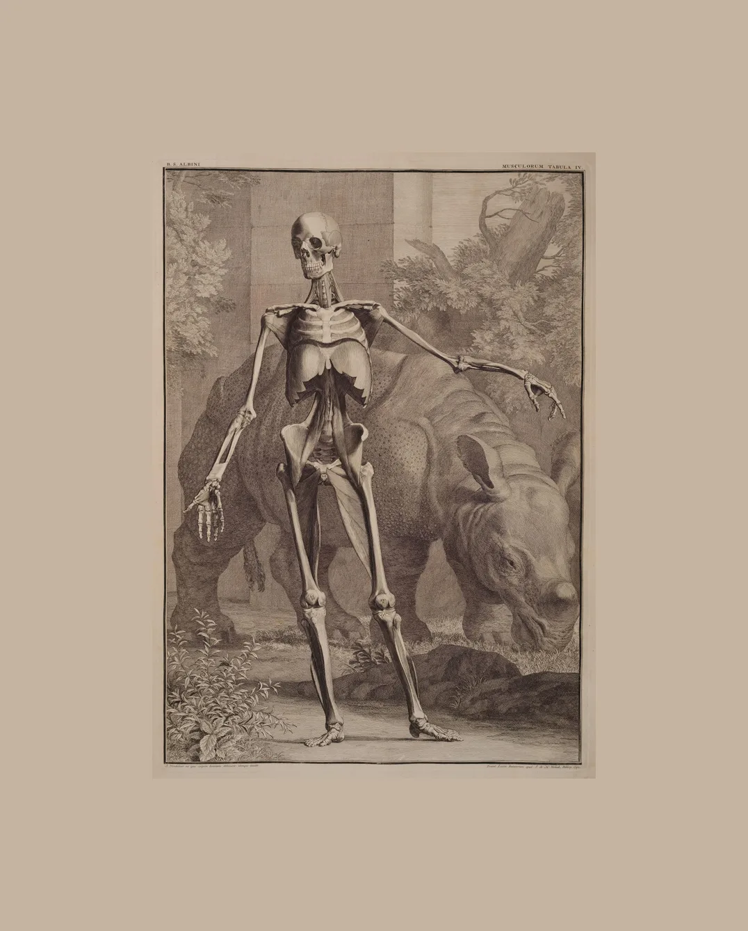
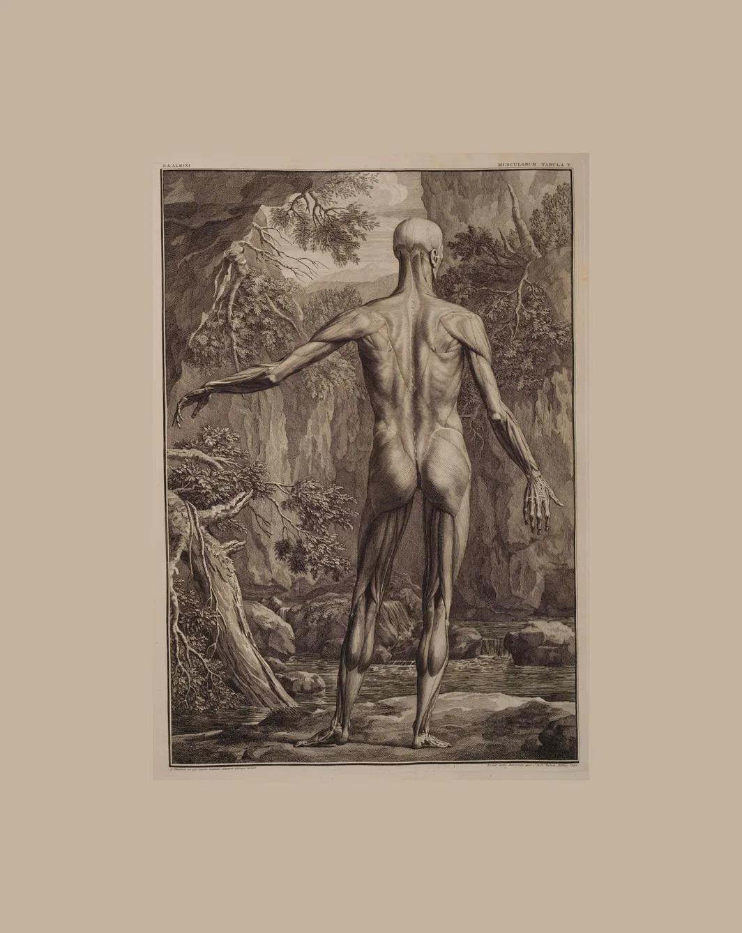
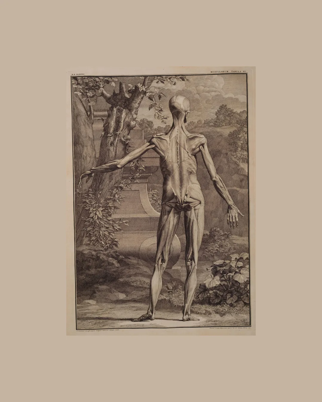
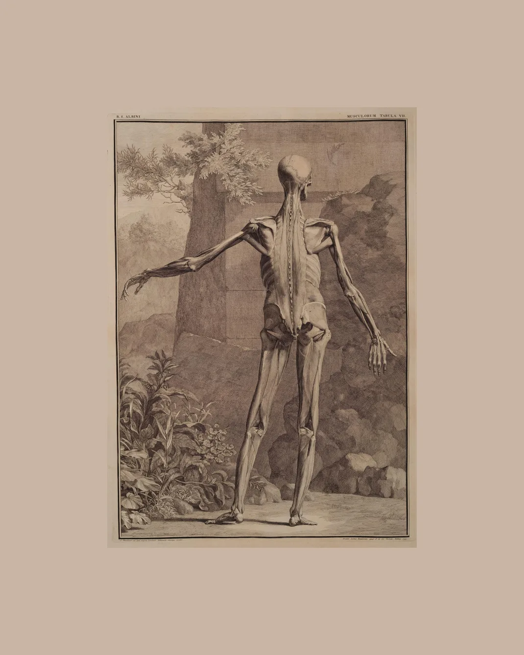
Albinus taught anatomy at Leiden for half a century. For this atlas he and the artist Jan Wandelaar drew the skeleton through a system of grids set at a distance from the body, to remove the distortion of perspective and show an ideal, measured figure.
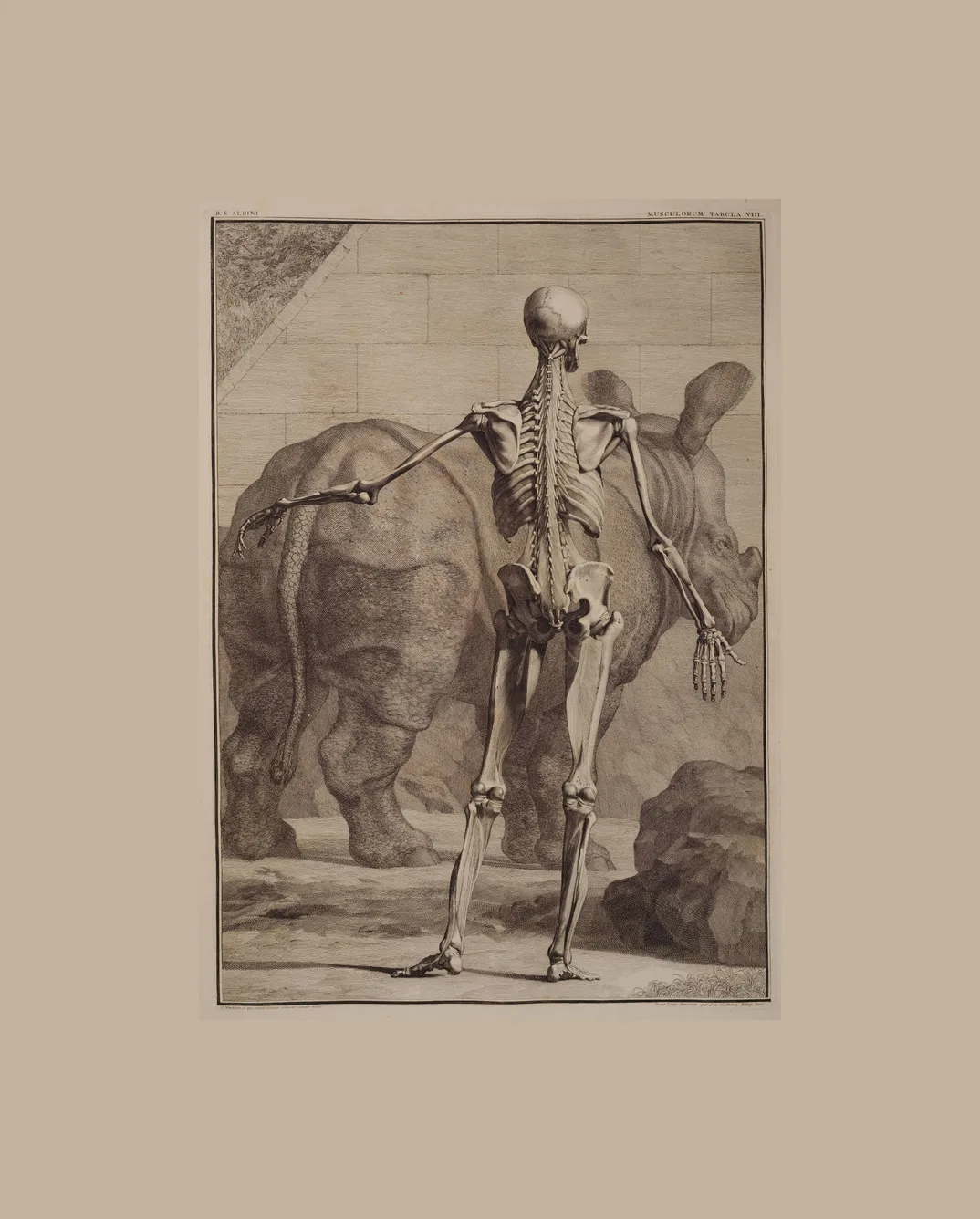
Wandelaar set the figures in gardens and ruins. Two of the muscle plates have a rhinoceros standing behind the figure: Clara, an Indian rhinoceros who arrived in Europe in 1741, became famous touring its cities, and was drawn from life by Wandelaar.
- Made by
- Engraved by Jan Wandelaar
- Published
- Leiden, 1747
- Medium
- Copper engraving
- On this page
- 18 plates
- See the original
- National Library of Medicine, Historical Anatomies on the Web
- 1830
Anatomical Plates
Published by Dr. Parker
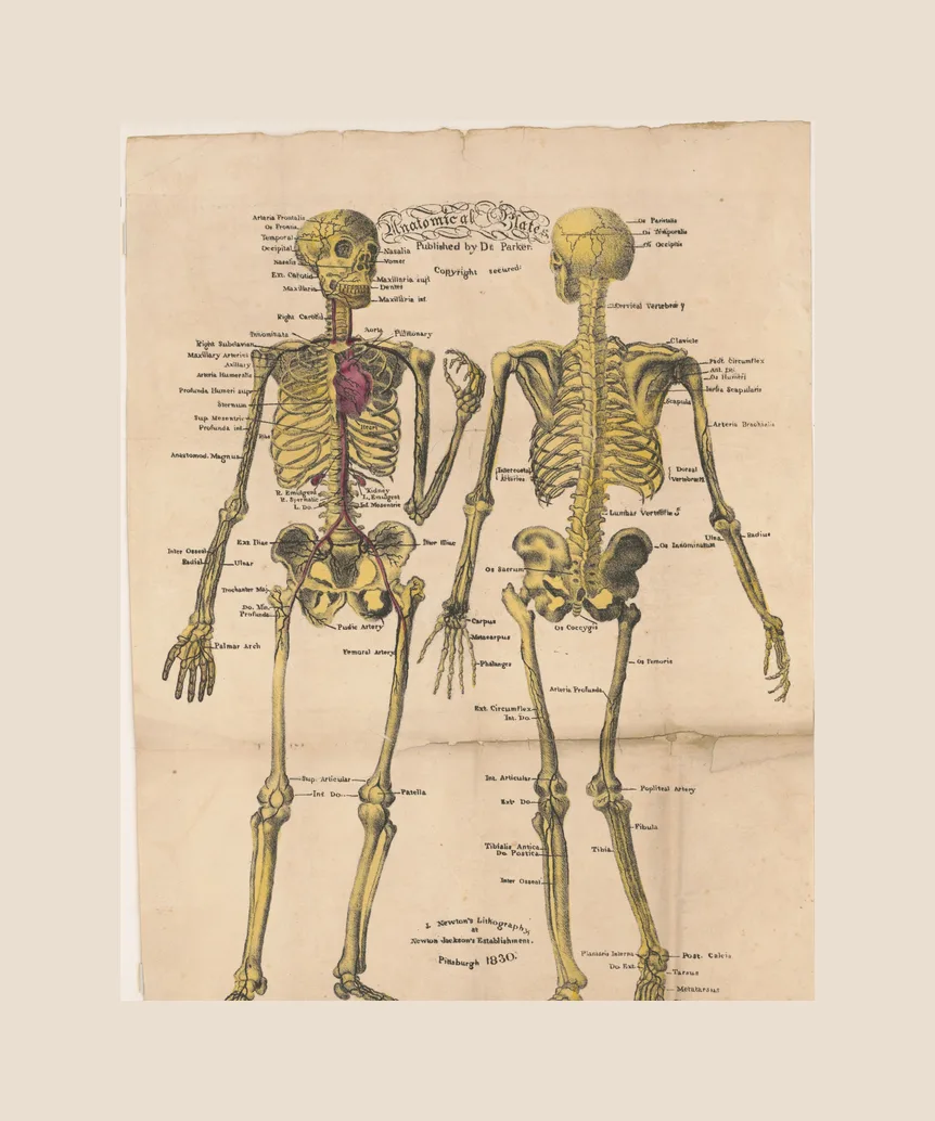
A teaching sheet of the 1830s: the skeleton with its arteries, front and back on one page, lettered for students. The copy we restored is held by the Library of Congress.
- Made by
- Lithographed by J. Newton
- Published
- United States, 1830
- Medium
- Lithograph
- On this page
- 1 plate
- See the original
- Library of Congress, Prints and Photographs
- 1904 / 1920
Atlas der deskriptiven Anatomie des Menschen
Johannes Sobotta (1869–1945)
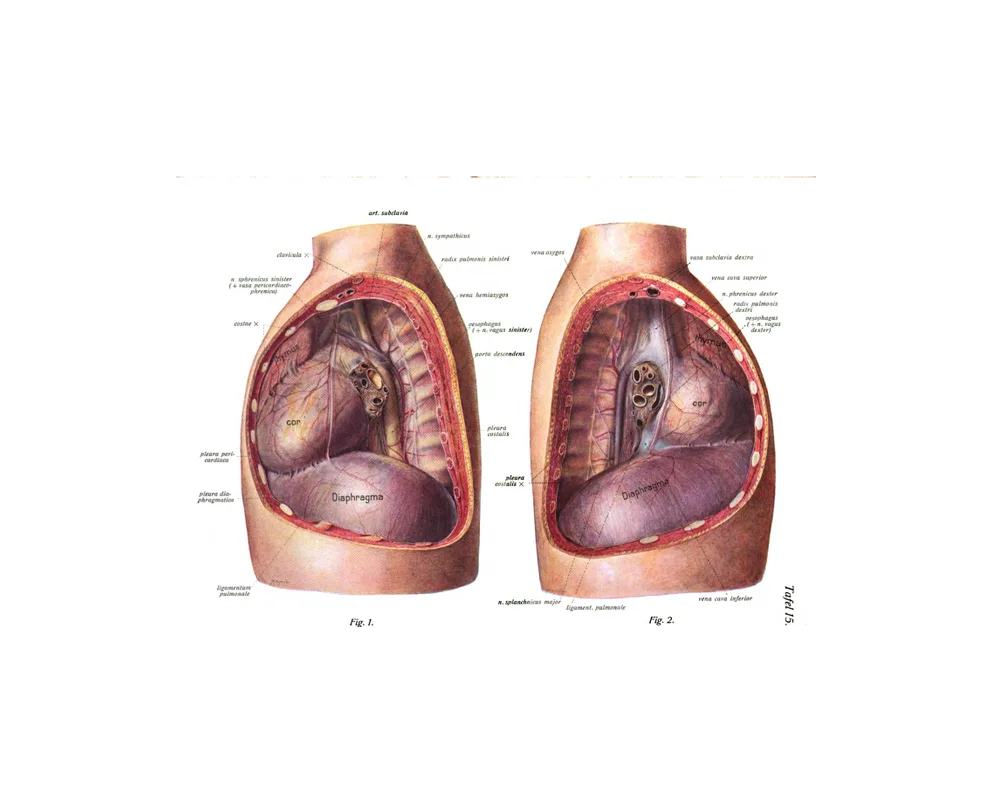
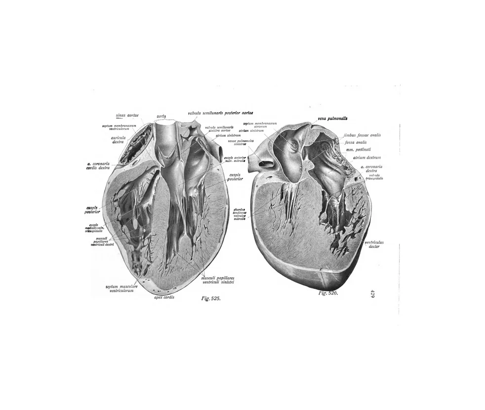
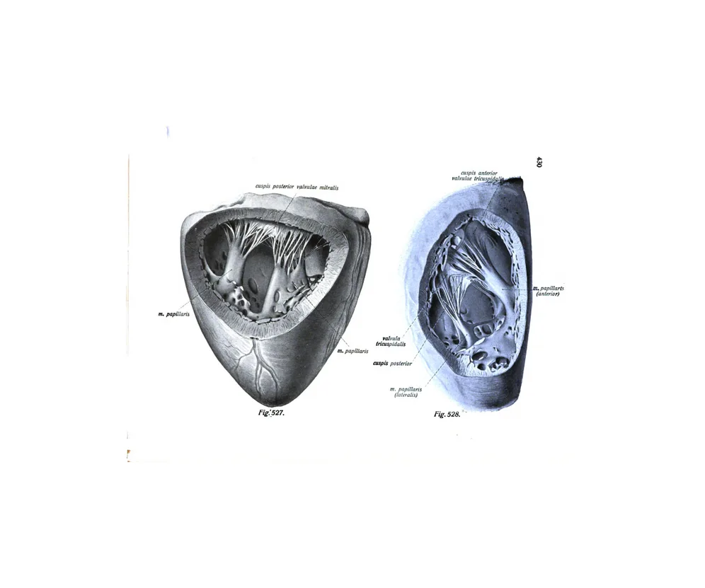
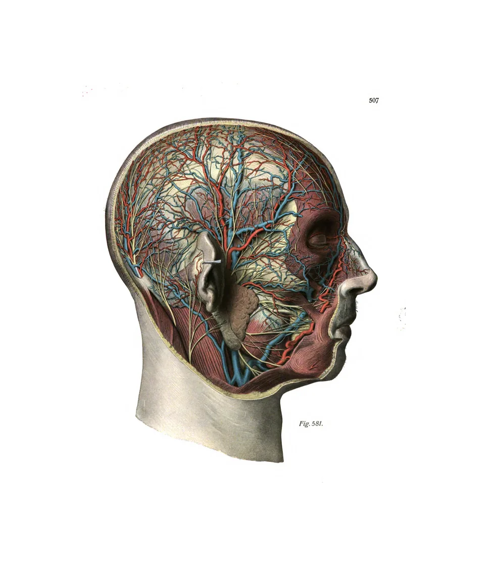
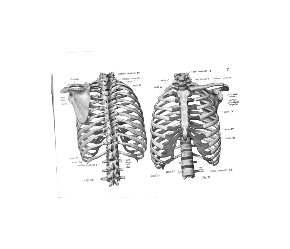
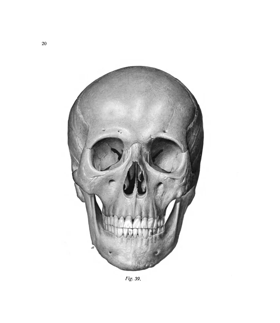
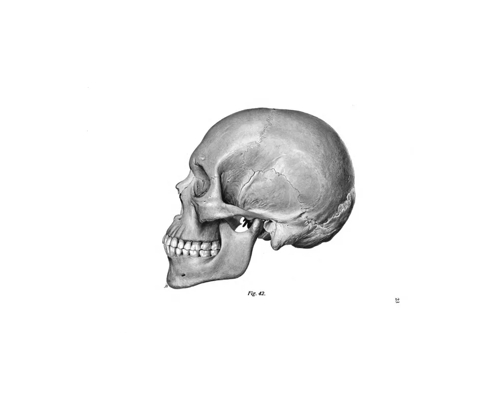
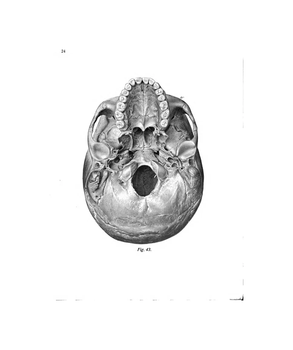
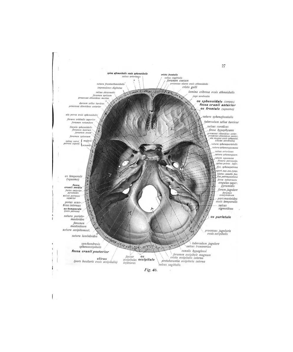
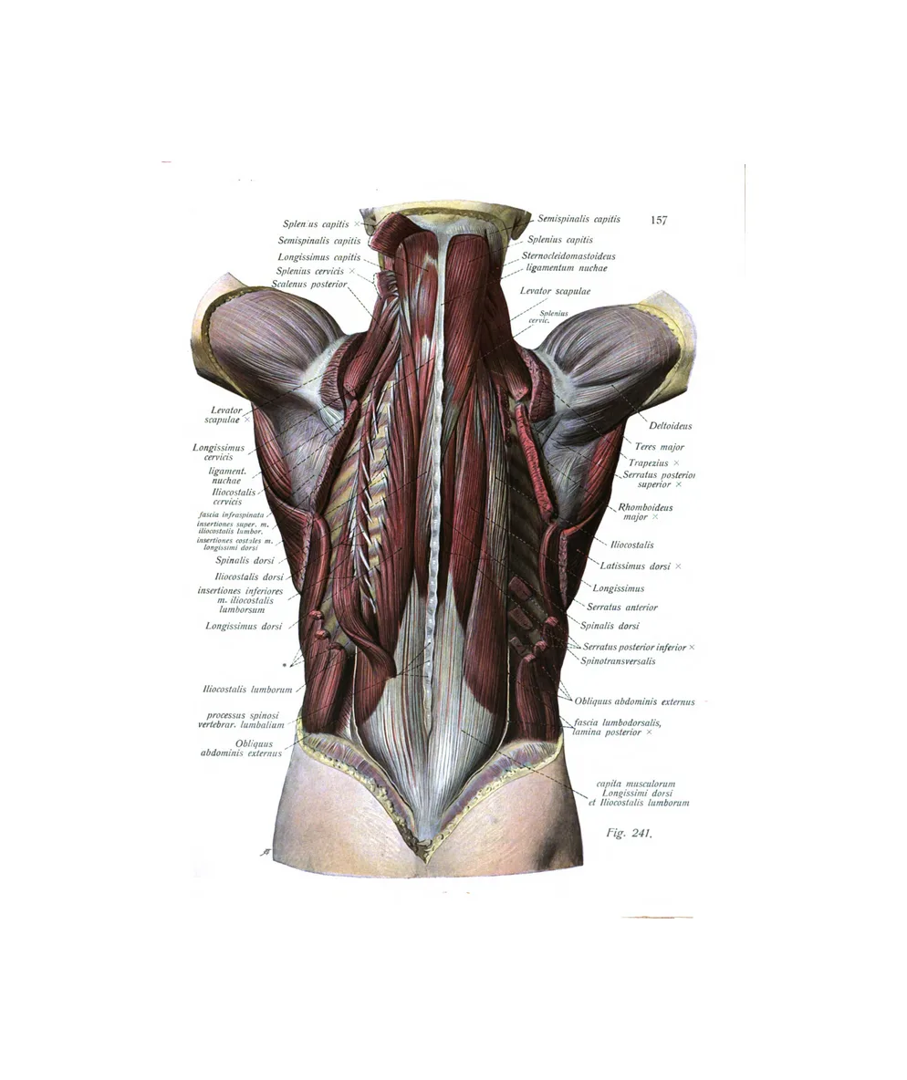
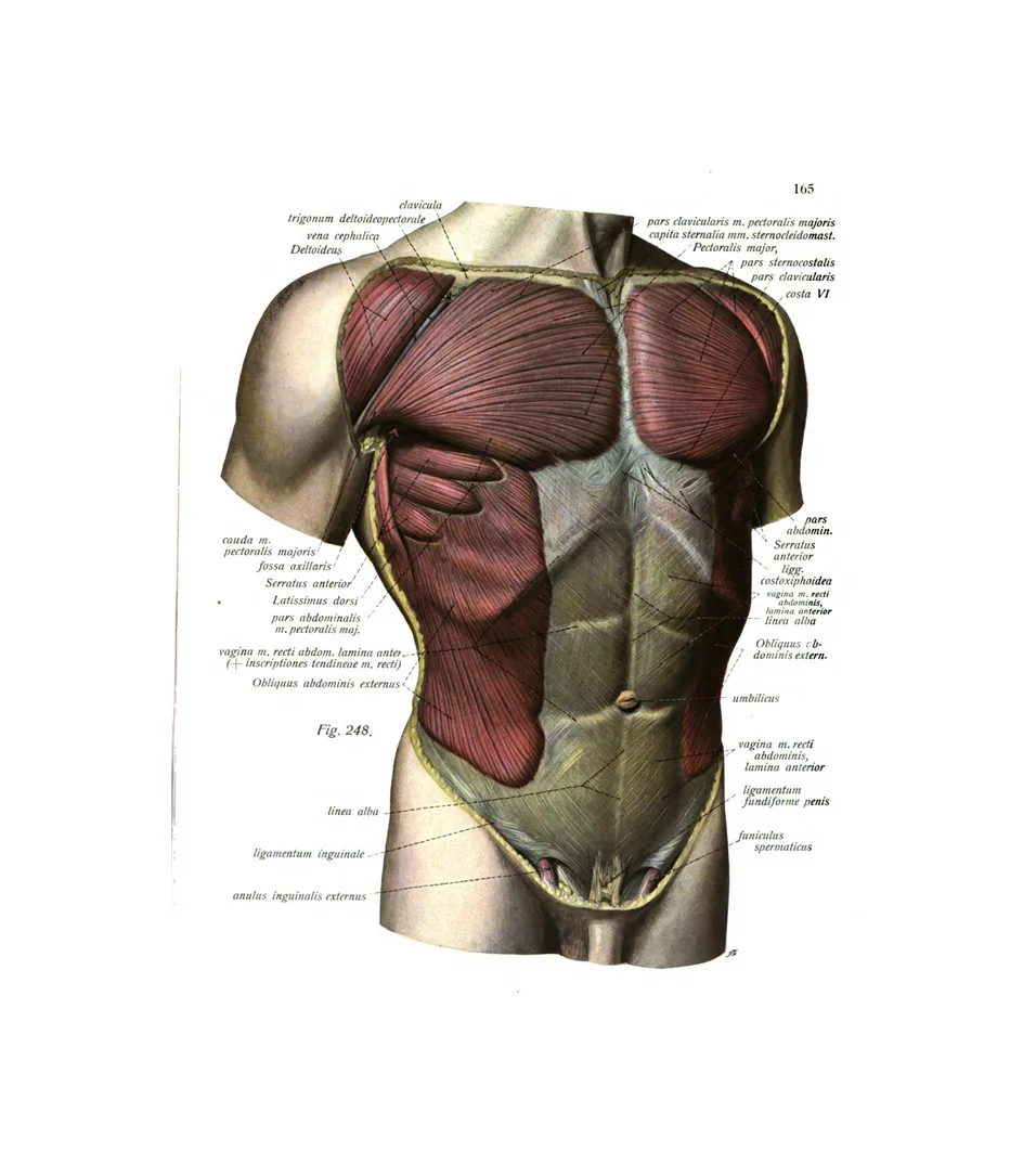
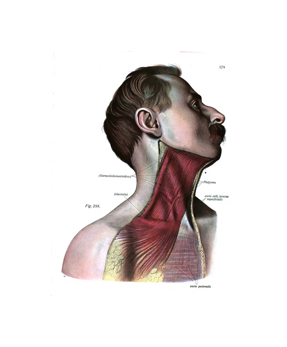
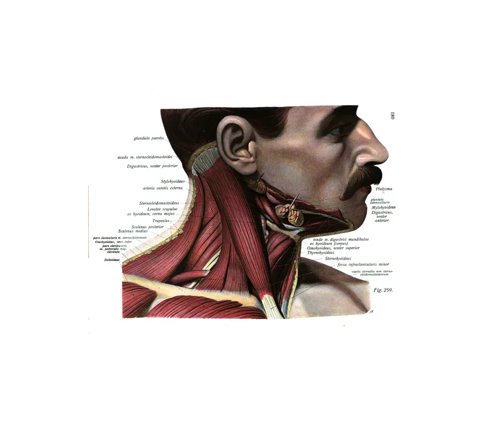
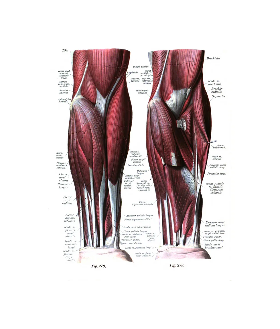
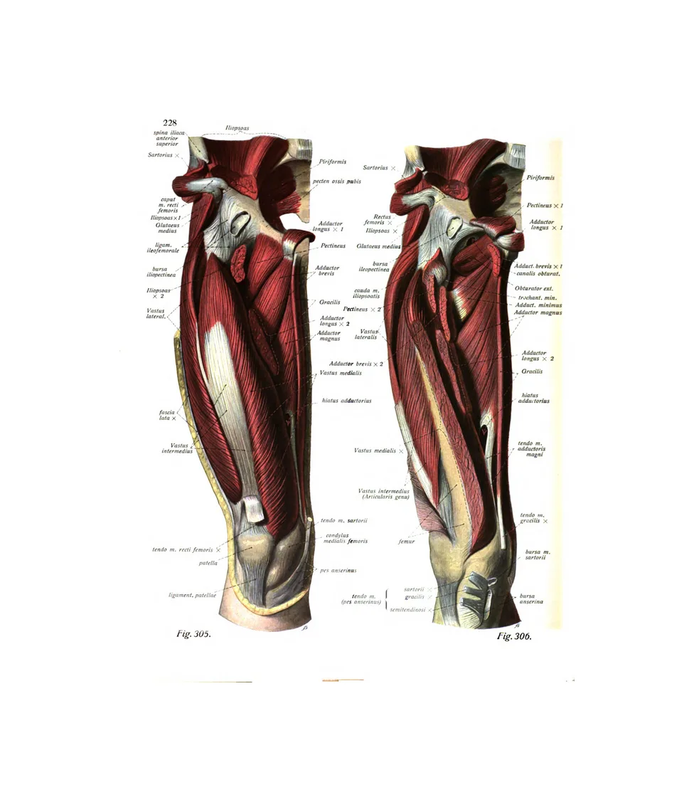
Sobotta’s atlas was made for medical students. It has been reissued in hundreds of editions and many languages, and it is still in print more than a century later. Karl Hajek’s colour plates show each part as it is found in dissection: the heart opened to its valves, the brain in section, the bones of the hand laid out one by one.
Where the older atlases show the whole body as a figure, Sobotta works part by part. That makes it the richest source here for the eye and ear, the teeth, the hand and the foot.
- Made by
- Illustrated by Karl Hajek and August Schmitson
- Published
- Munich: J. F. Lehmann, first published 1904; our plates are from the 1919–1920 printing
- Medium
- Colour lithograph
- On this page
- 72 plates
- See the original
- Internet Archive, vol. I (1919) · vol. II (1920) · vol. III (1920)
By body part
The heart
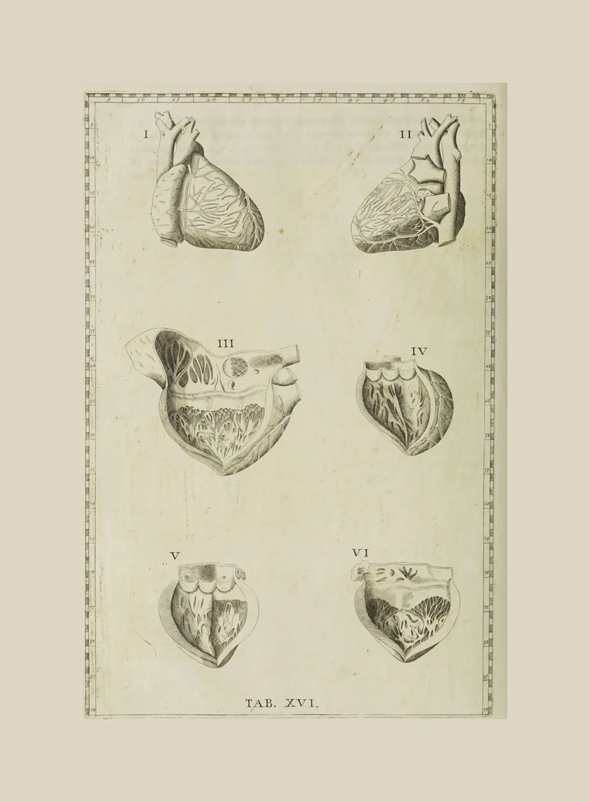
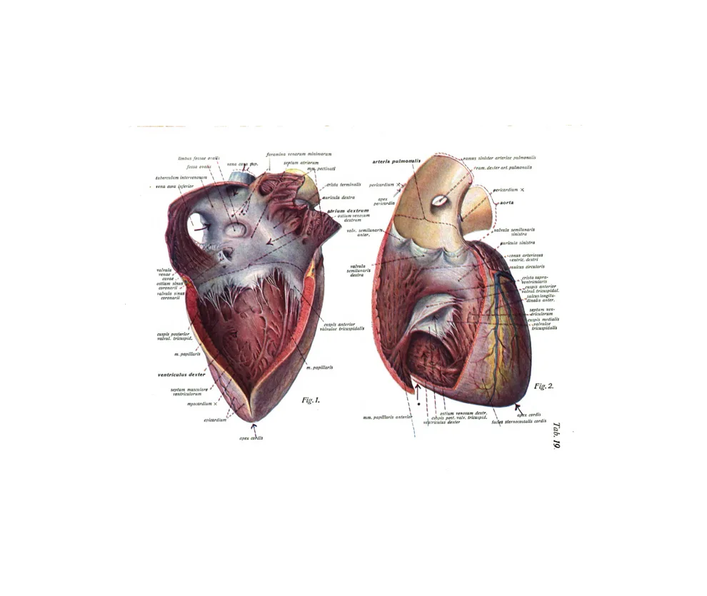
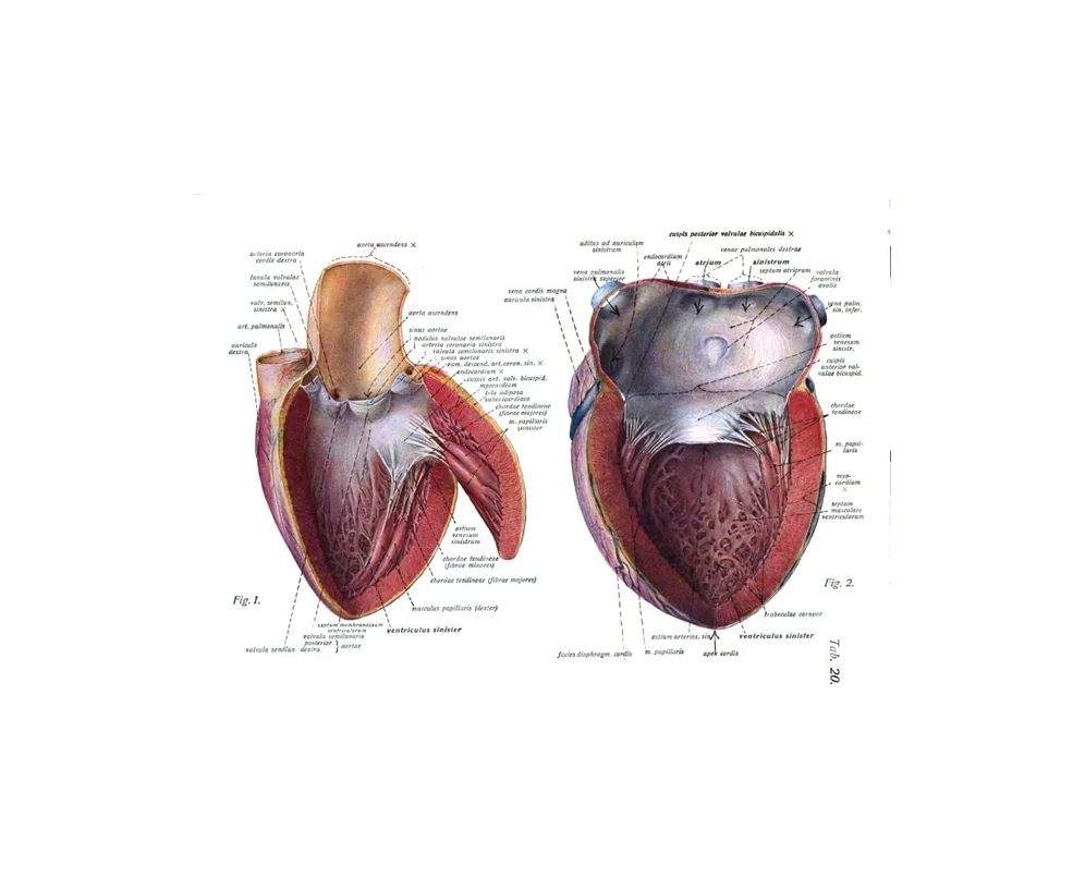
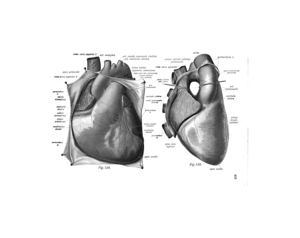
7 plates · Eustachi, SobottaEustachi shows the heart in six views on one plate. Sobotta’s colour plates open it chamber by chamber, to the valves.
The lungs and chest
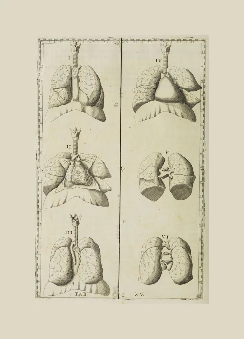
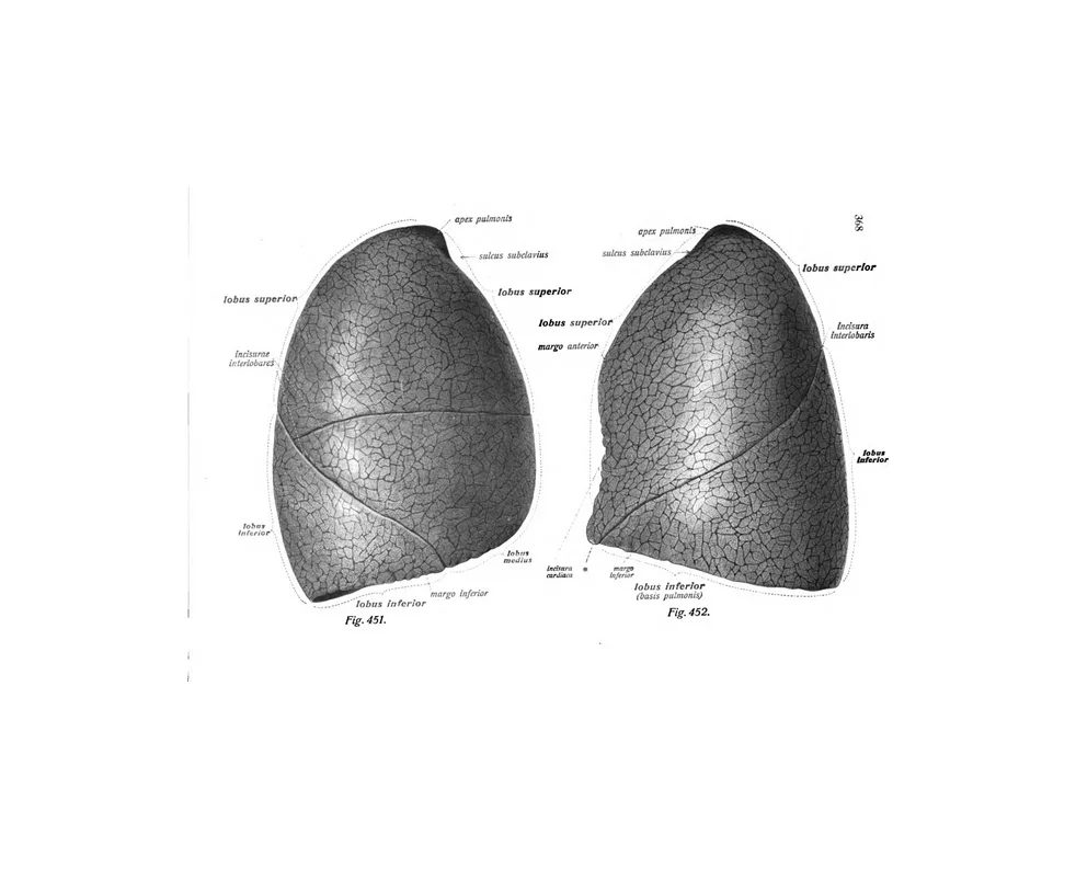
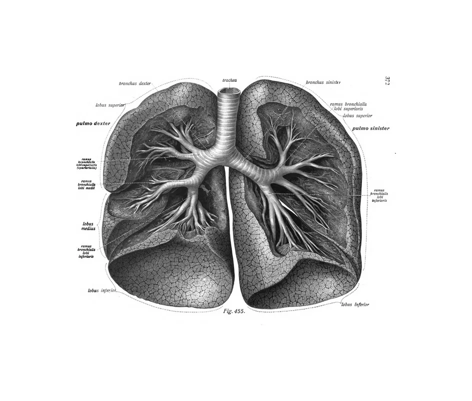
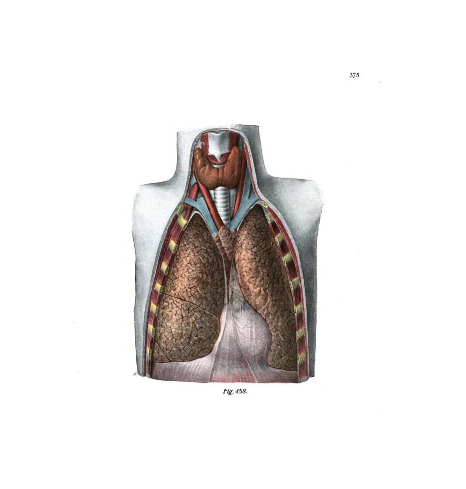
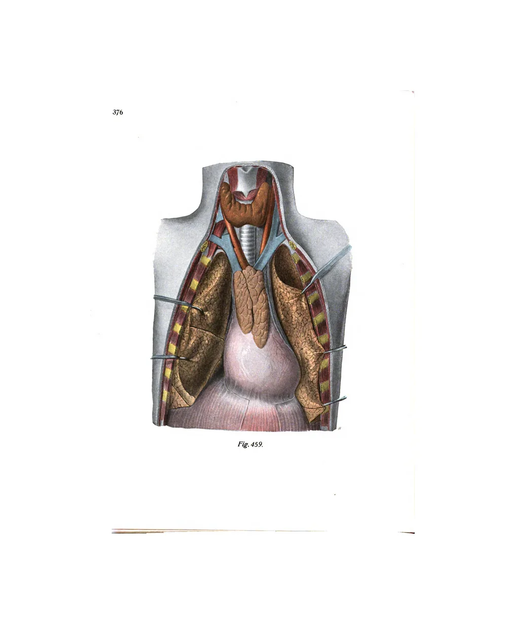
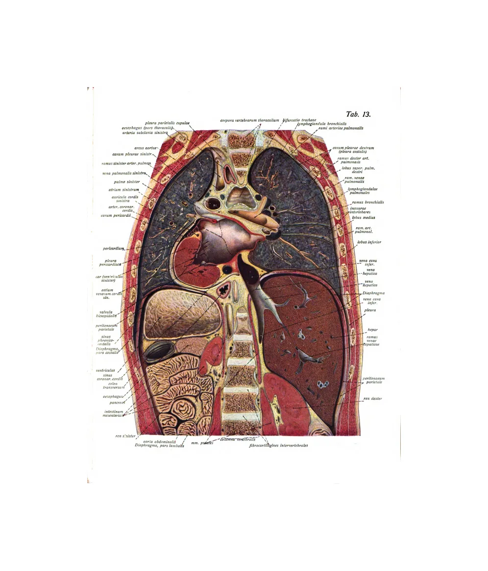
6 plates · Eustachi, SobottaThe lungs, the windpipe and its branches, and the chest opened or cut in section to show how the heart and lungs sit together.
The brain and nerves
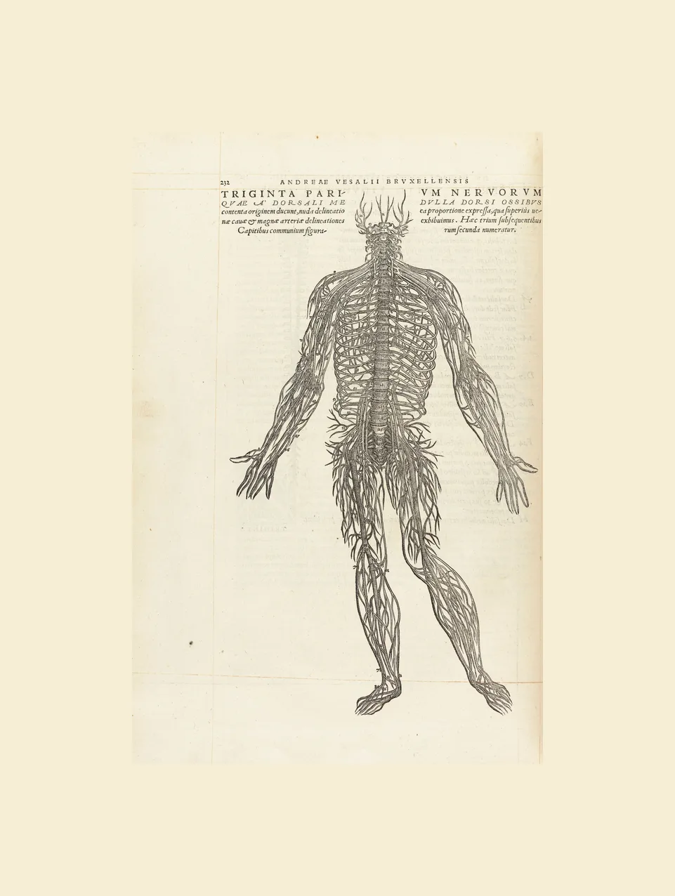
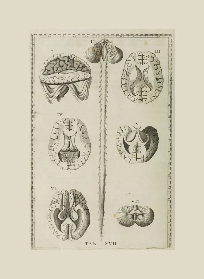
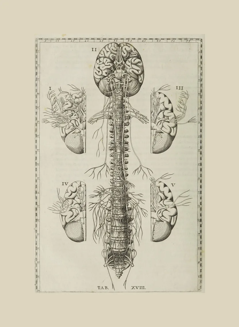
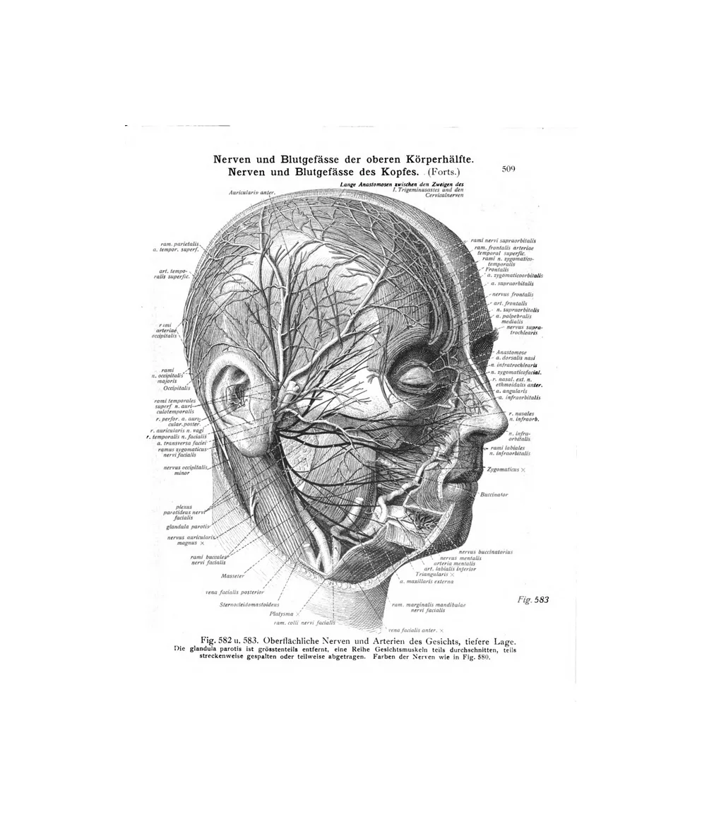
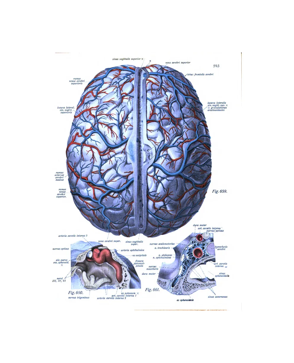
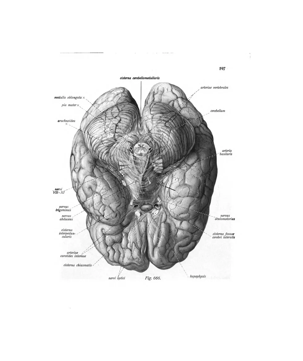
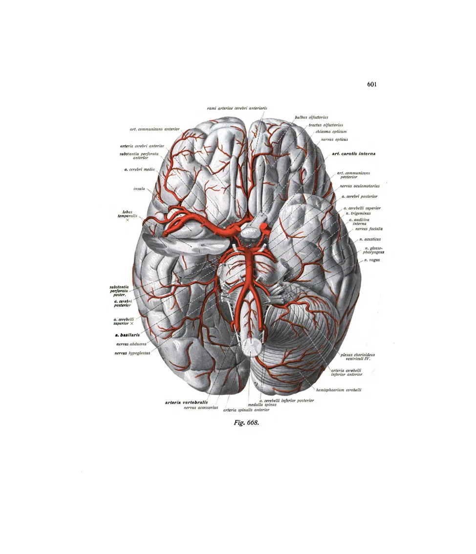
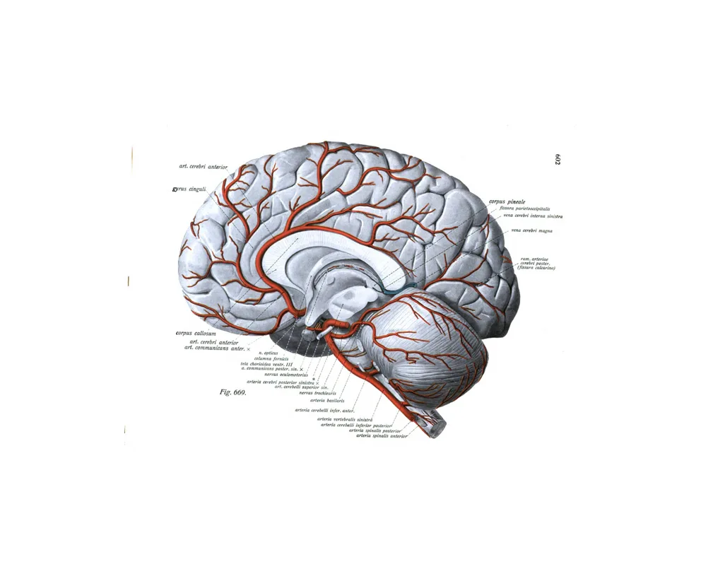
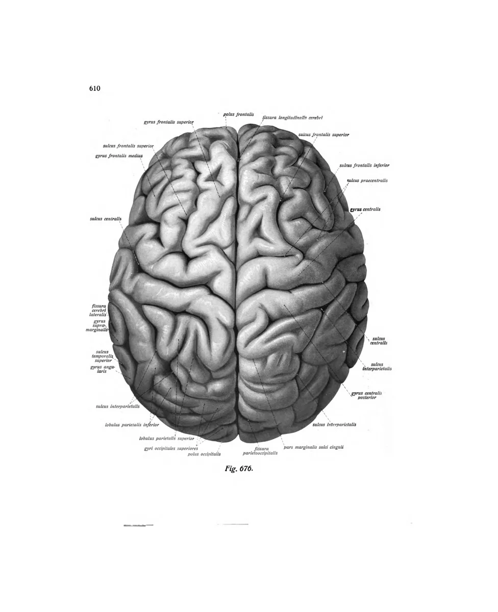
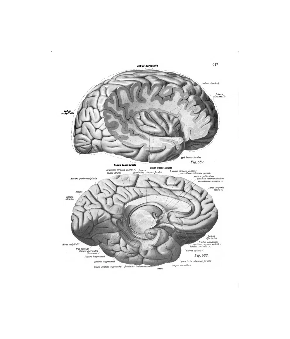
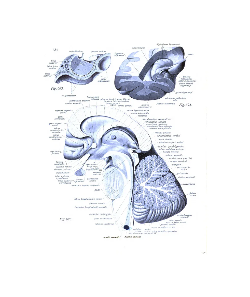
13 plates · Vesalius, Eustachi, SobottaFrom Vesalius’s figure of the thirty pairs of nerves to Sobotta’s brain in section, with its arteries and veins.
The eye and ear
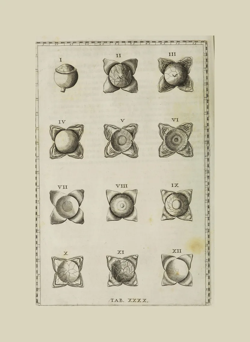
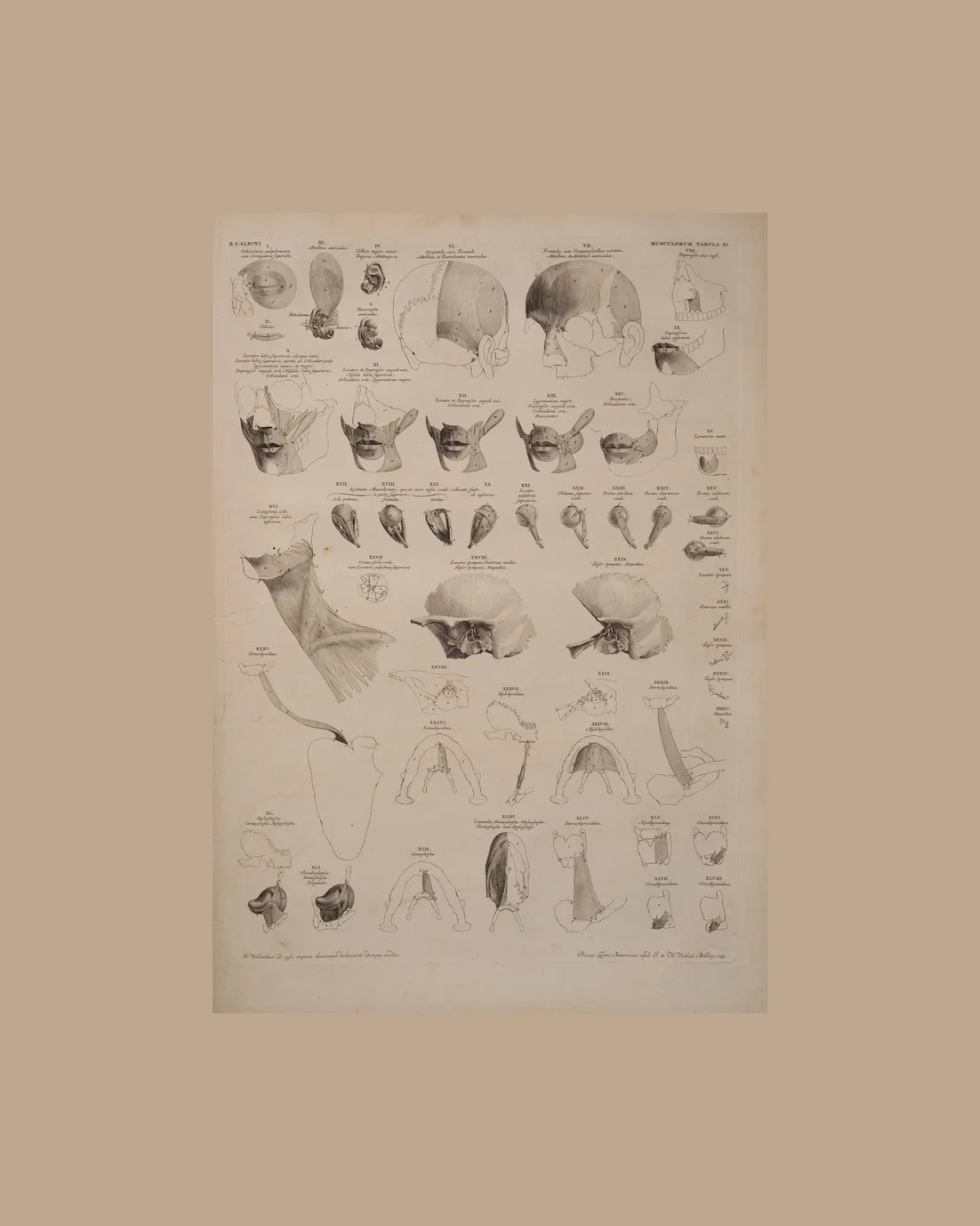
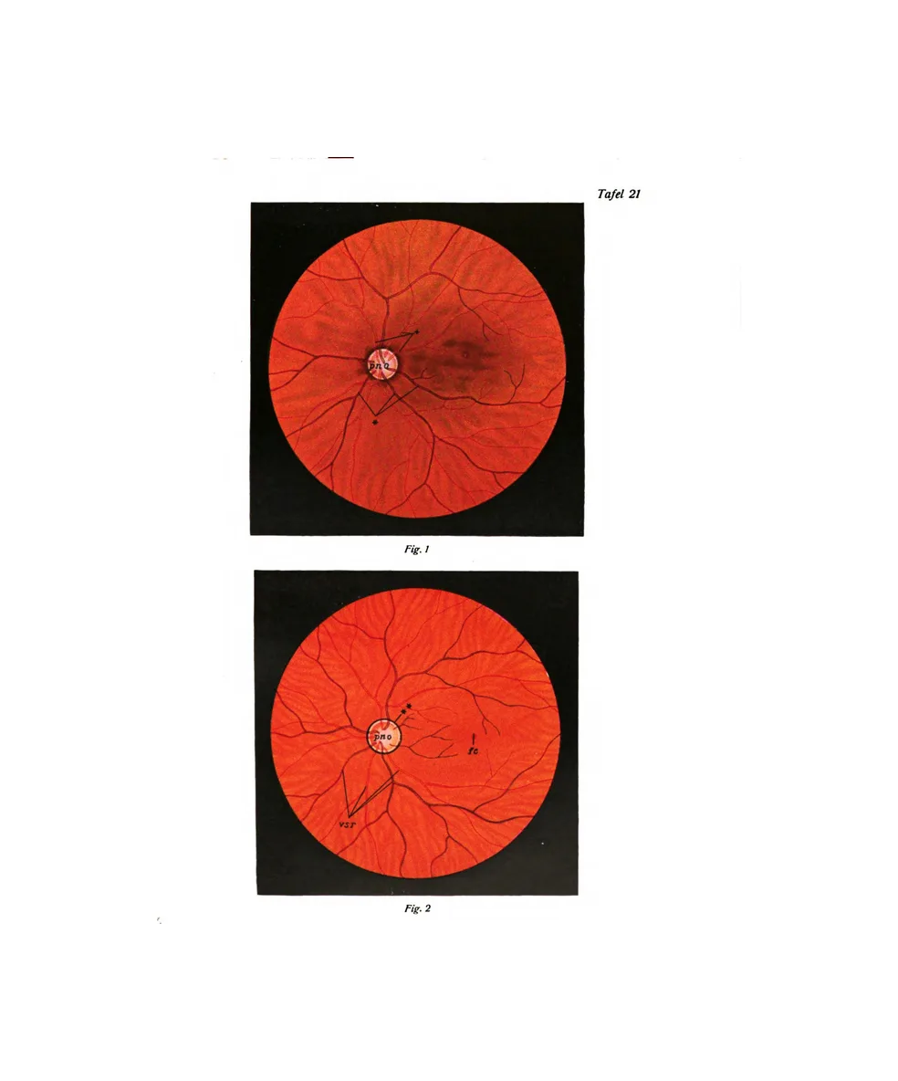
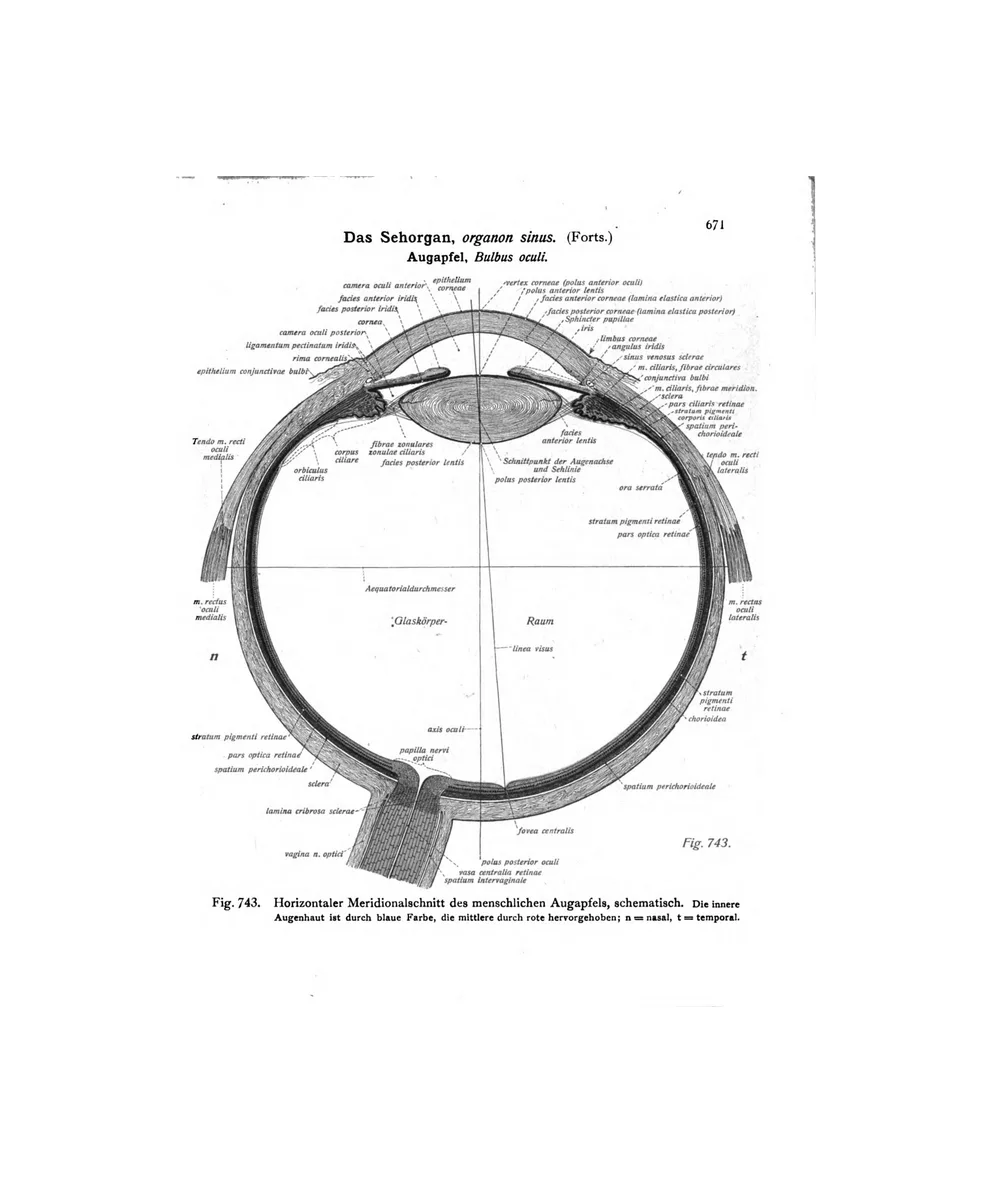
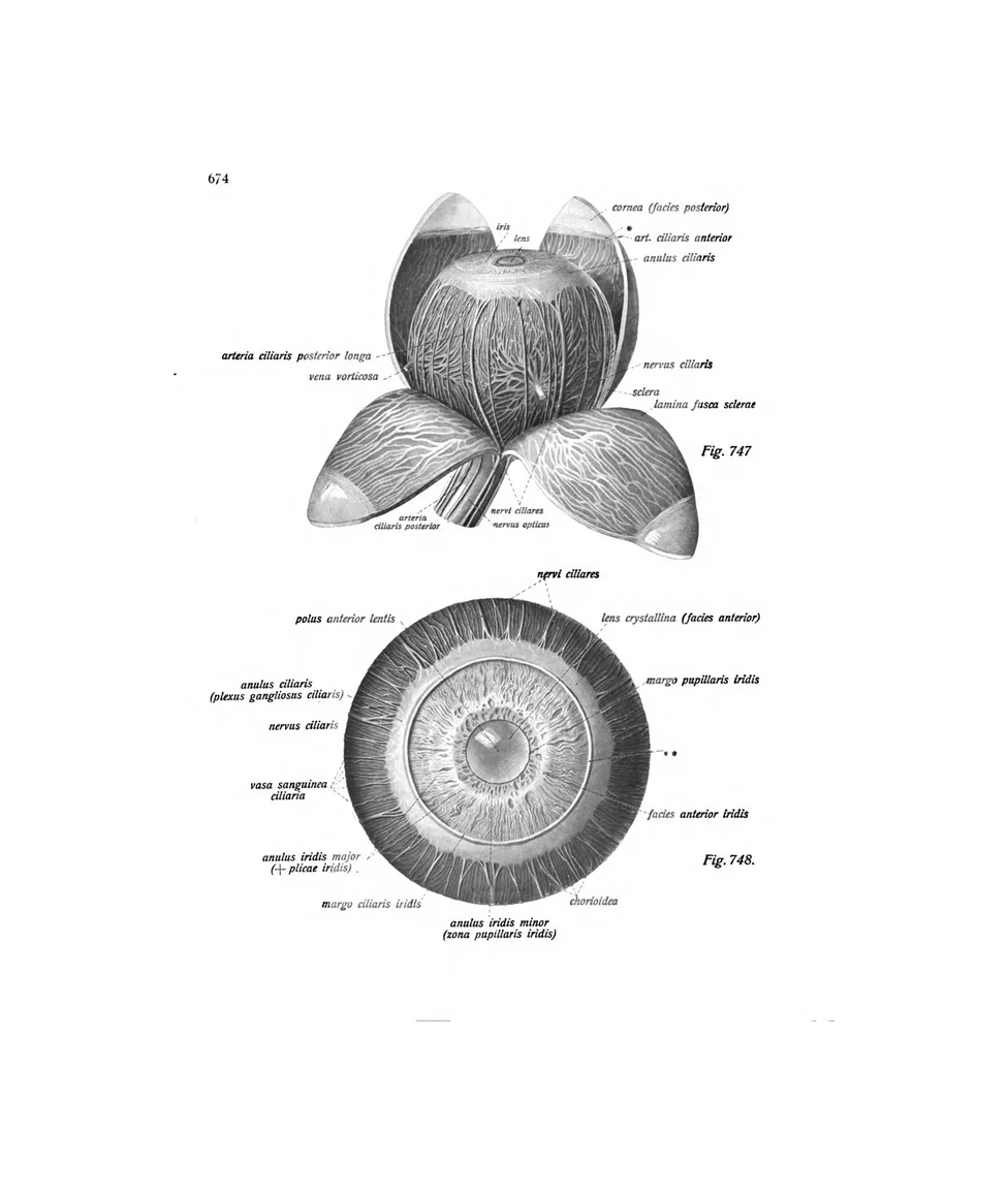
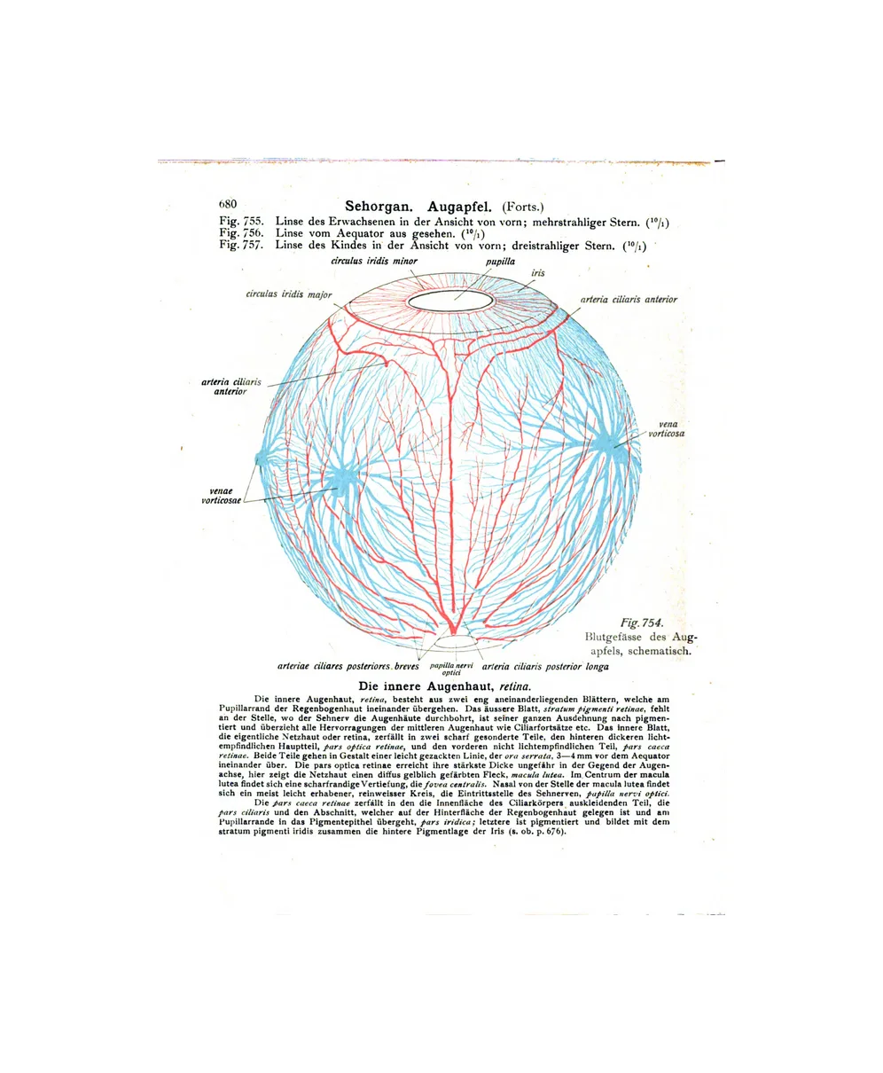
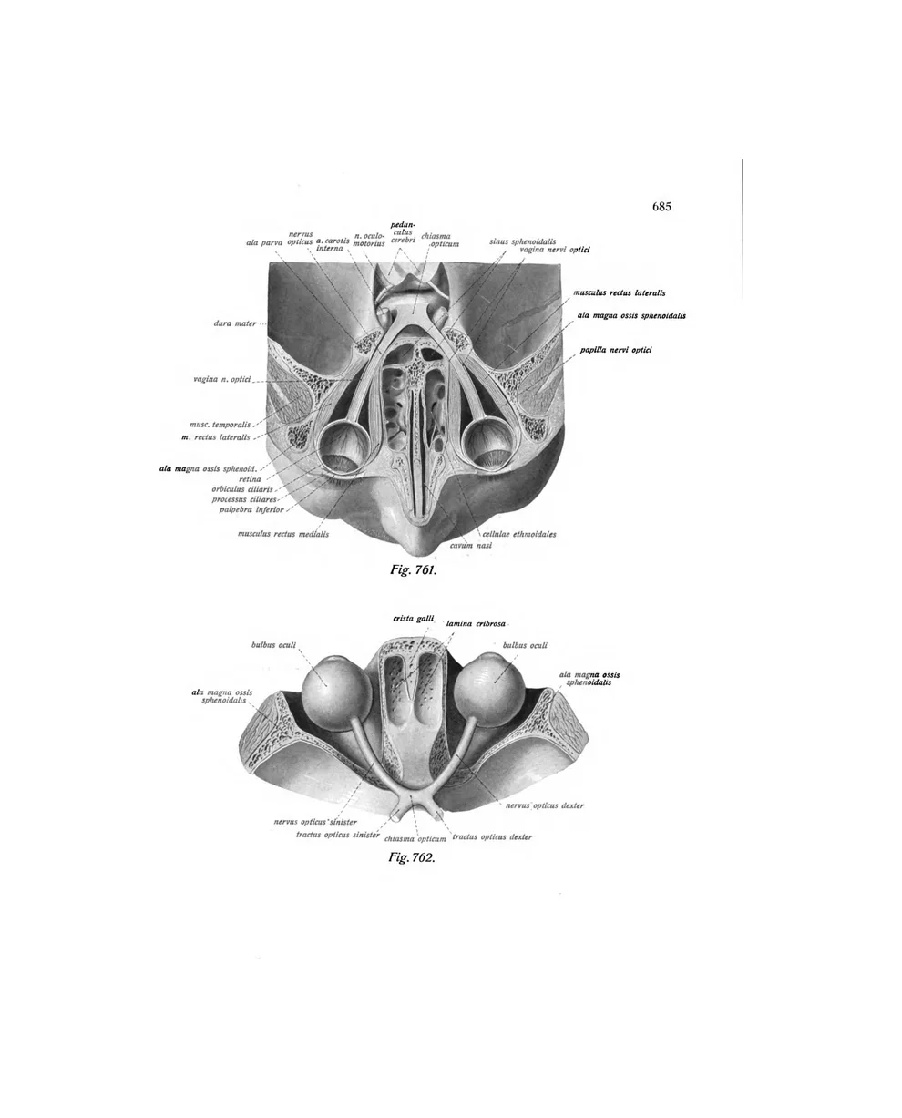
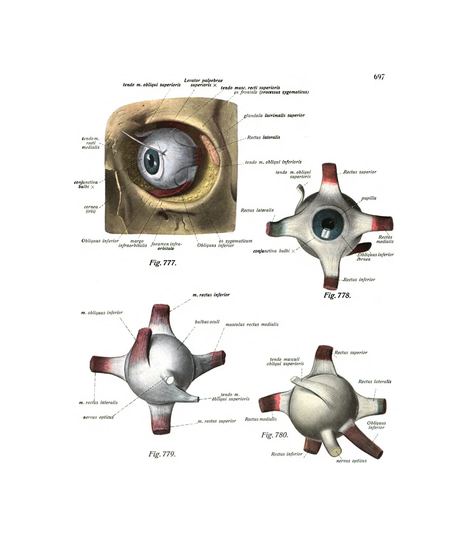
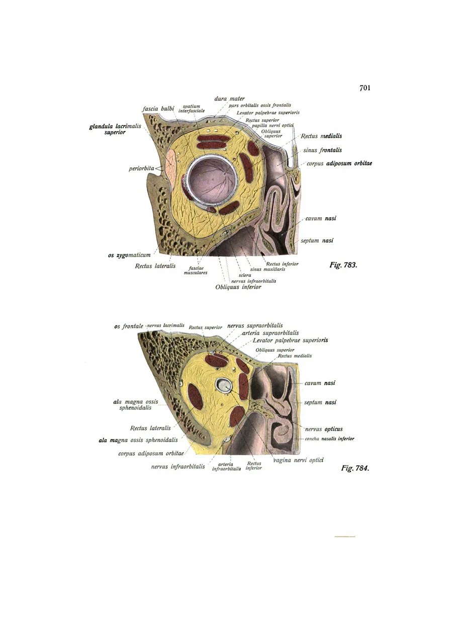
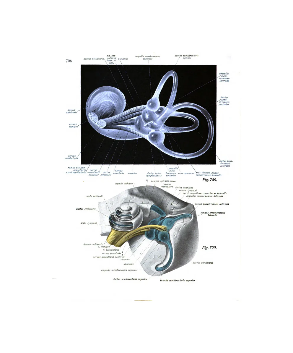
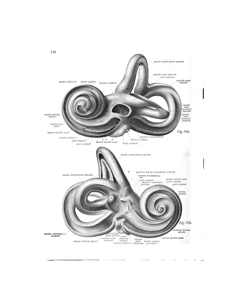
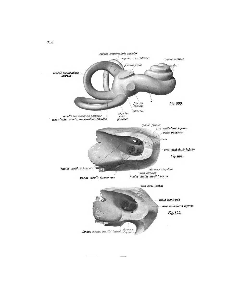
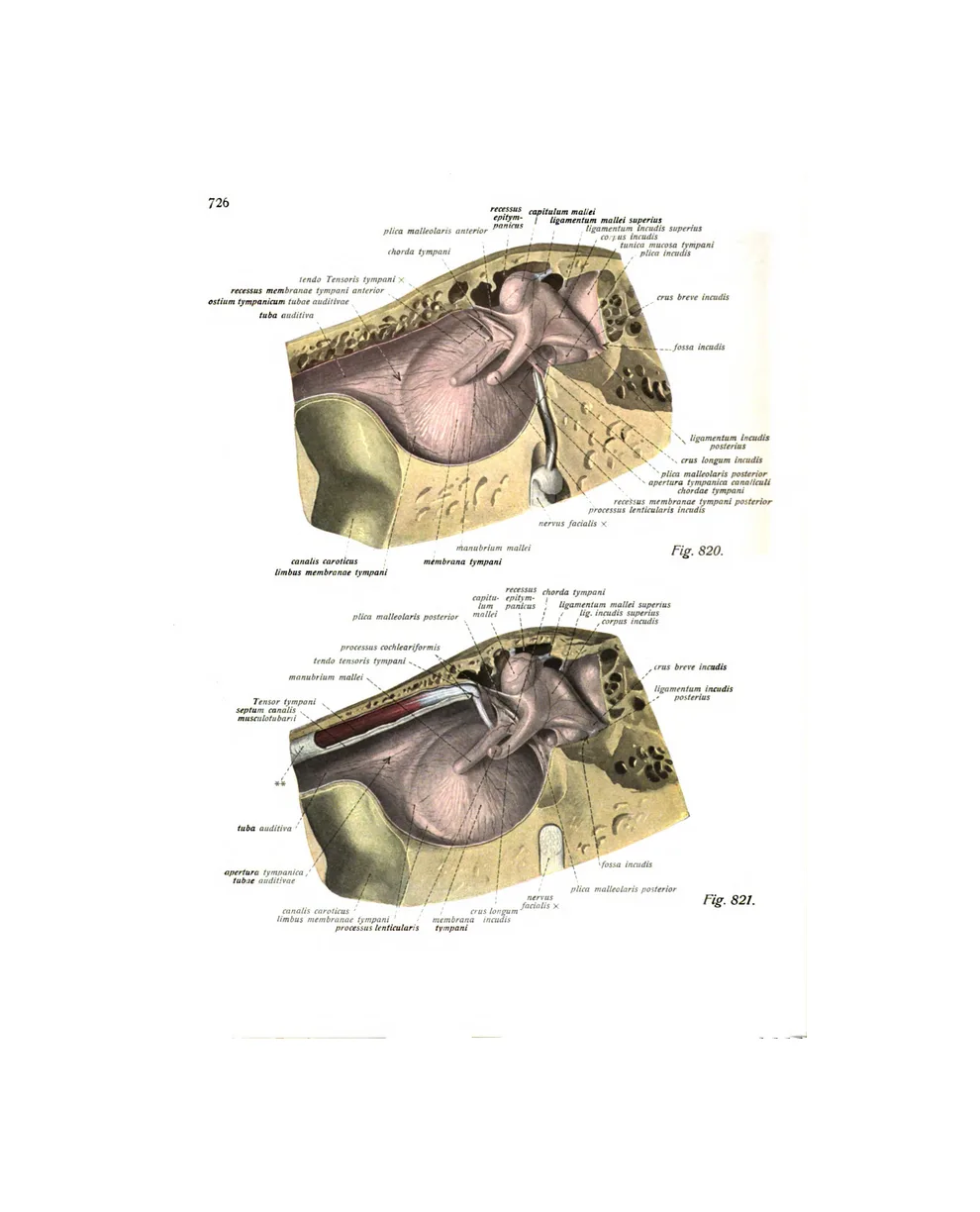
13 plates · Eustachi, Albinus, SobottaEustachi’s twelve dissections of the eye, and Sobotta’s eye in its orbit, the retina, and the inner ear in colour.
The teeth and jaw
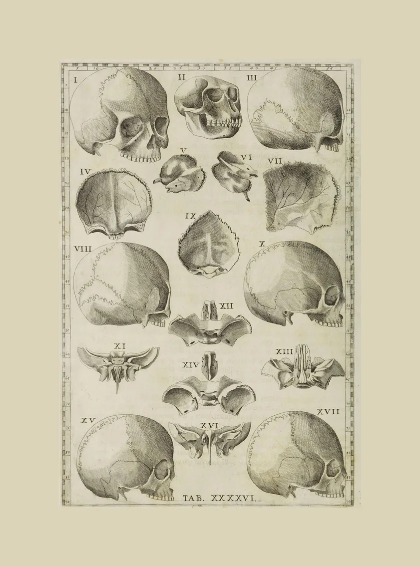
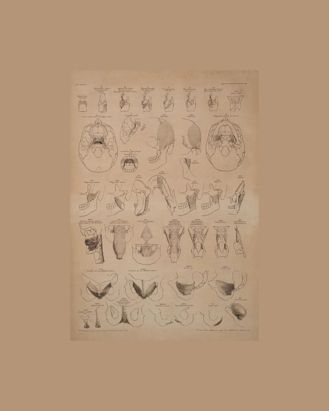
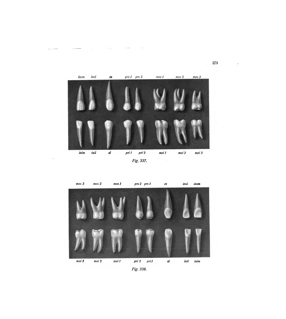
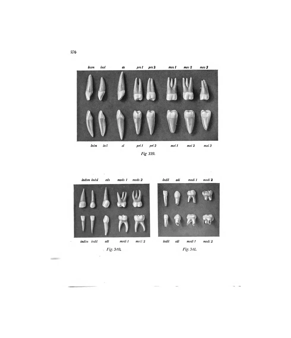
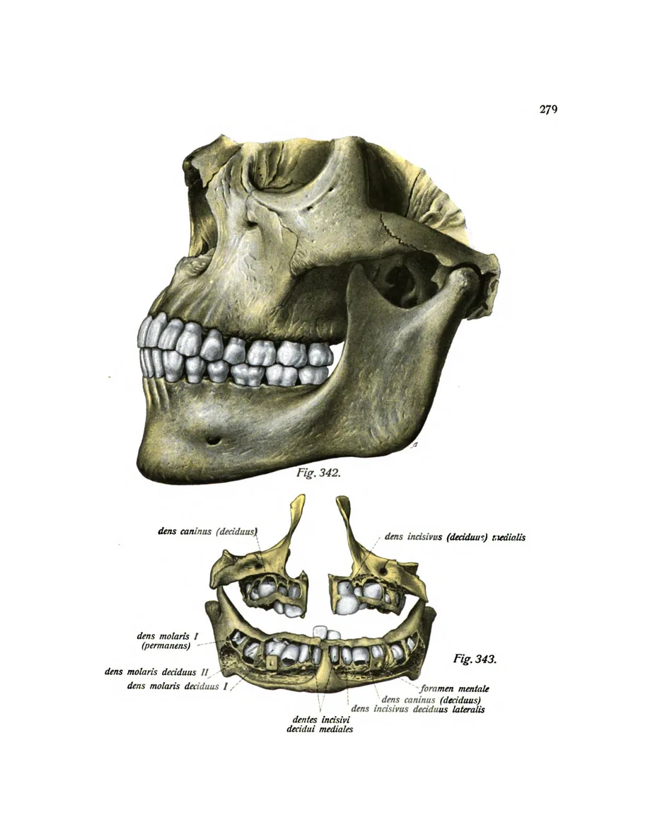
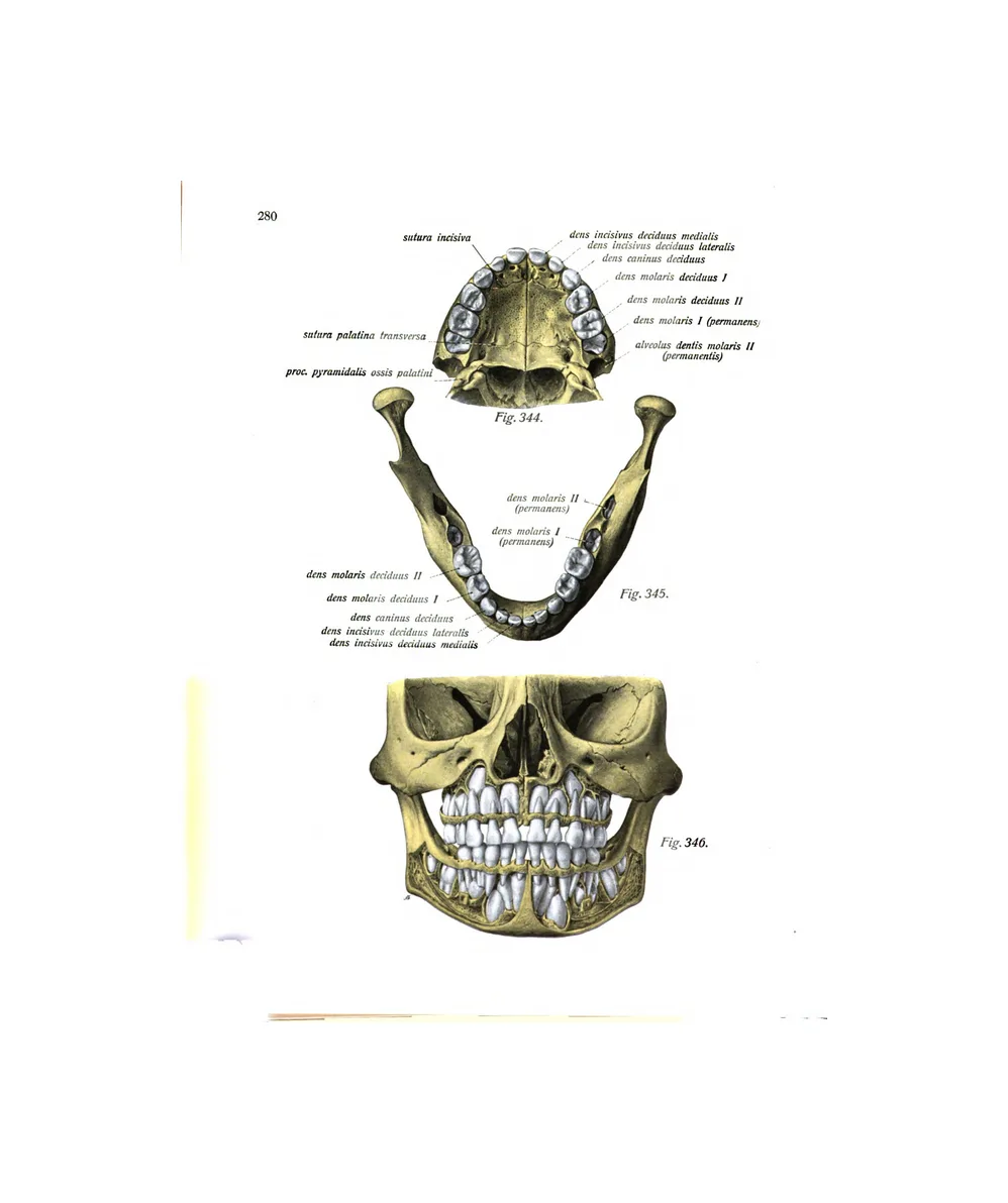
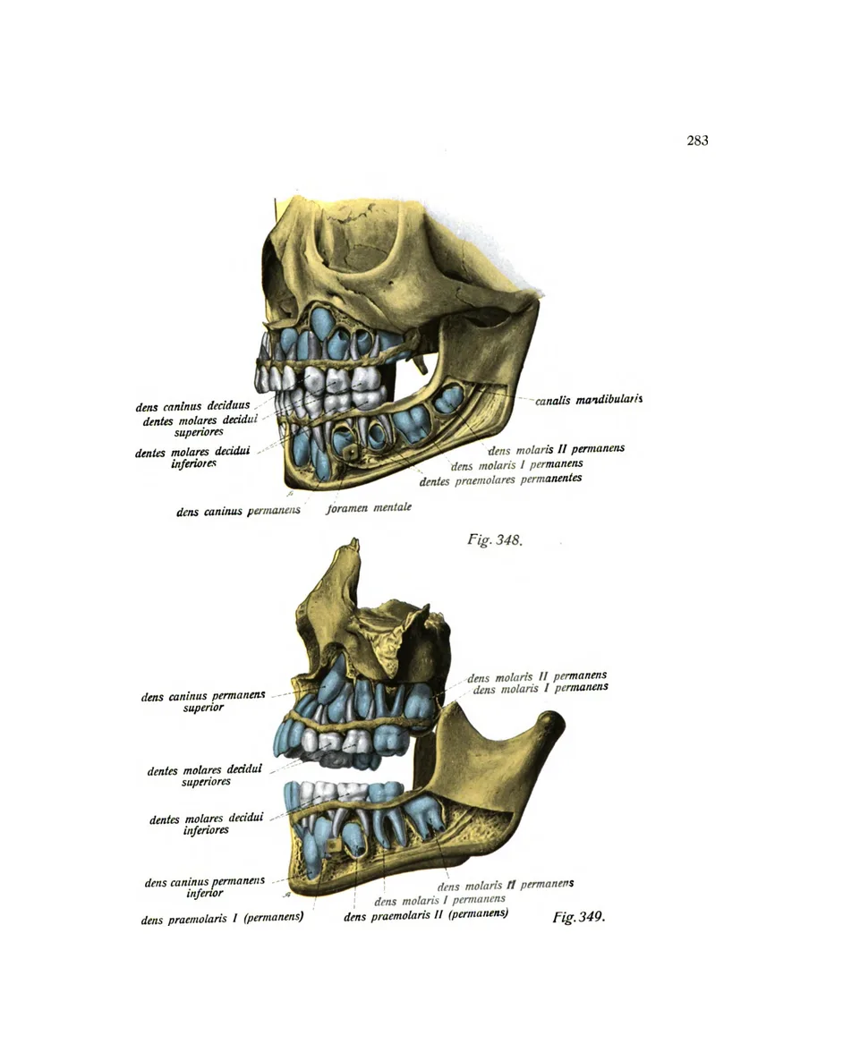
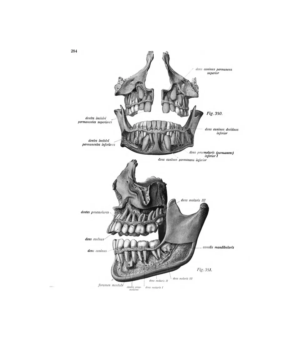
8 plates · Eustachi, Albinus, SobottaThe skull in seventeen views, the permanent and milk teeth, and children’s skulls showing the teeth still forming.
The hand
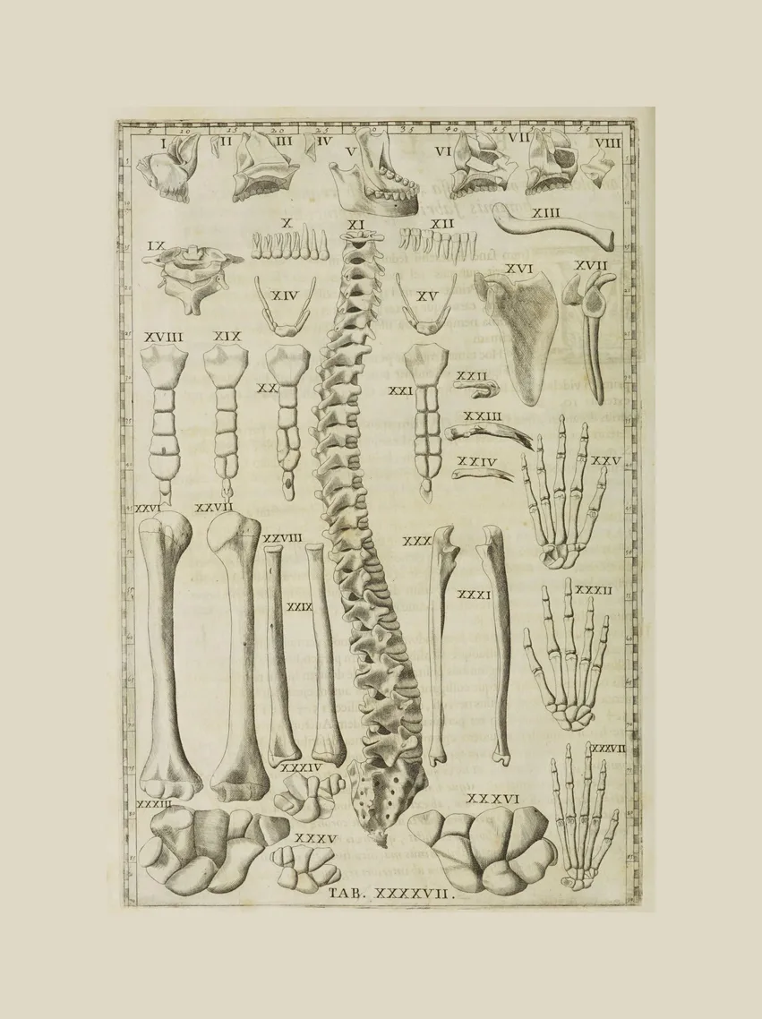
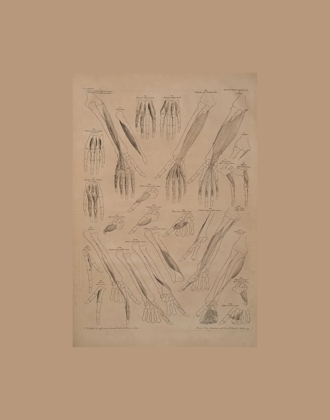
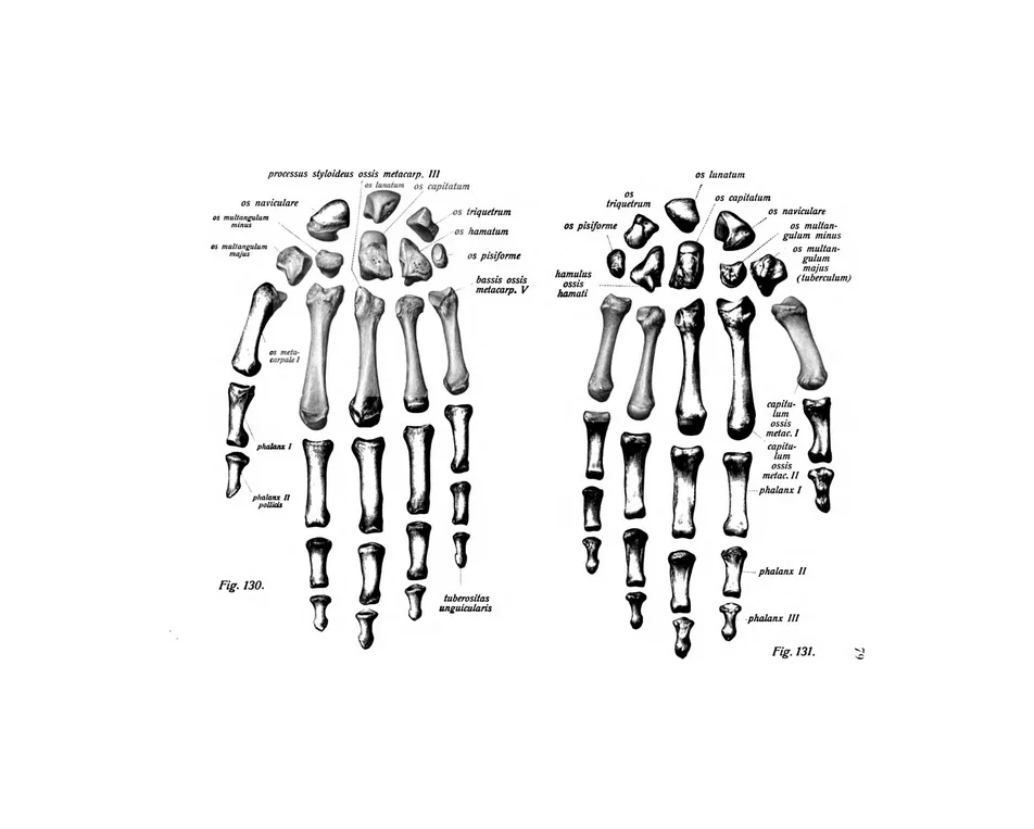
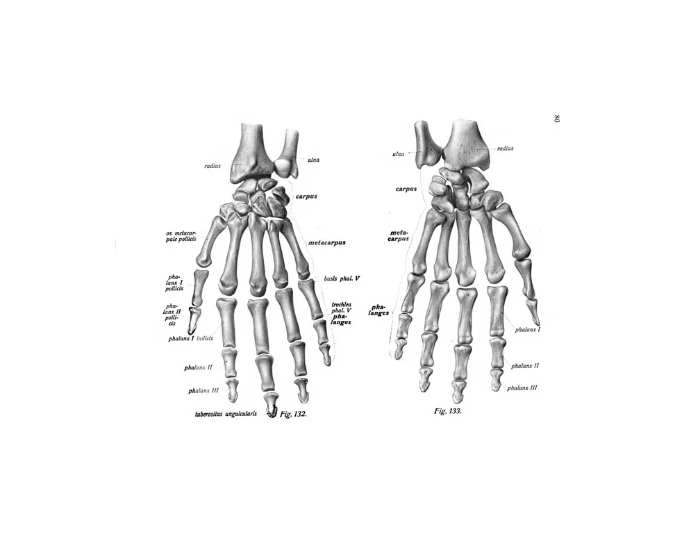
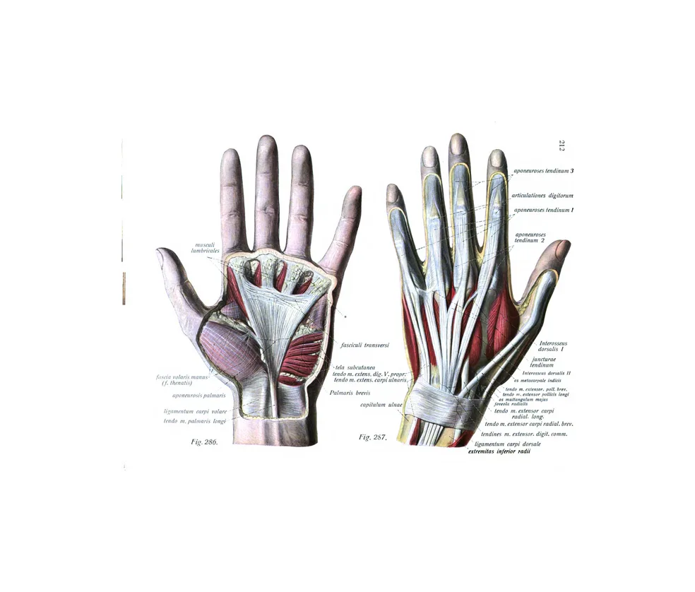
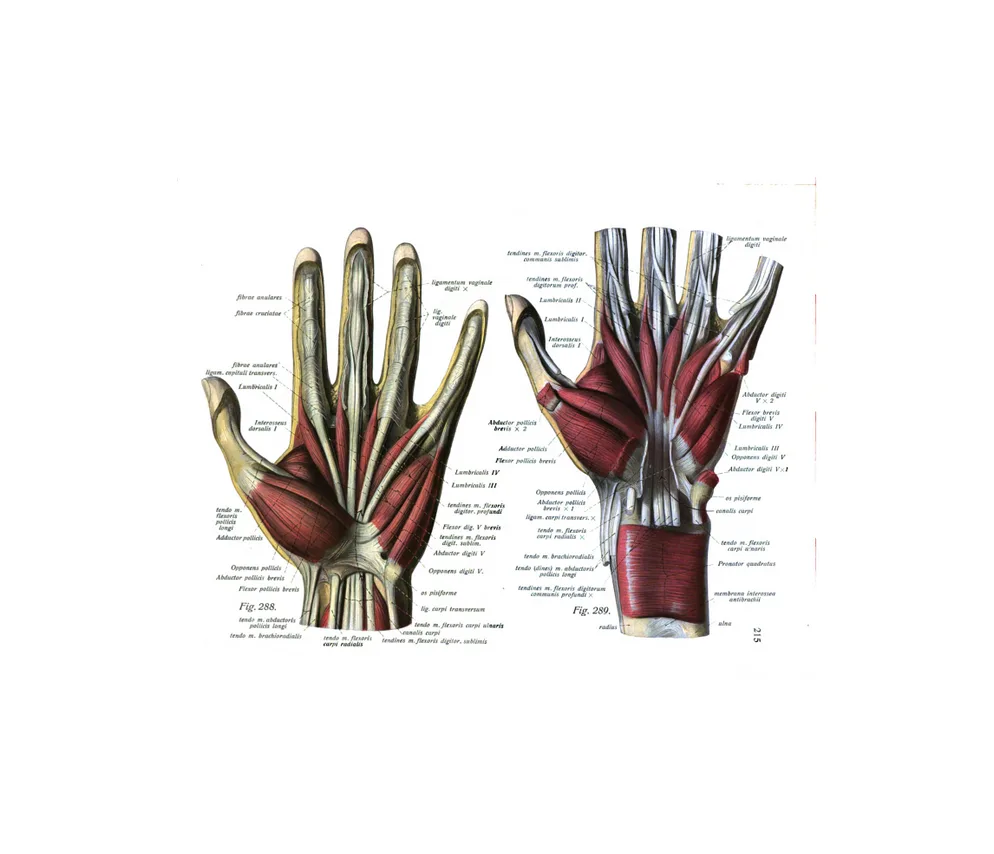
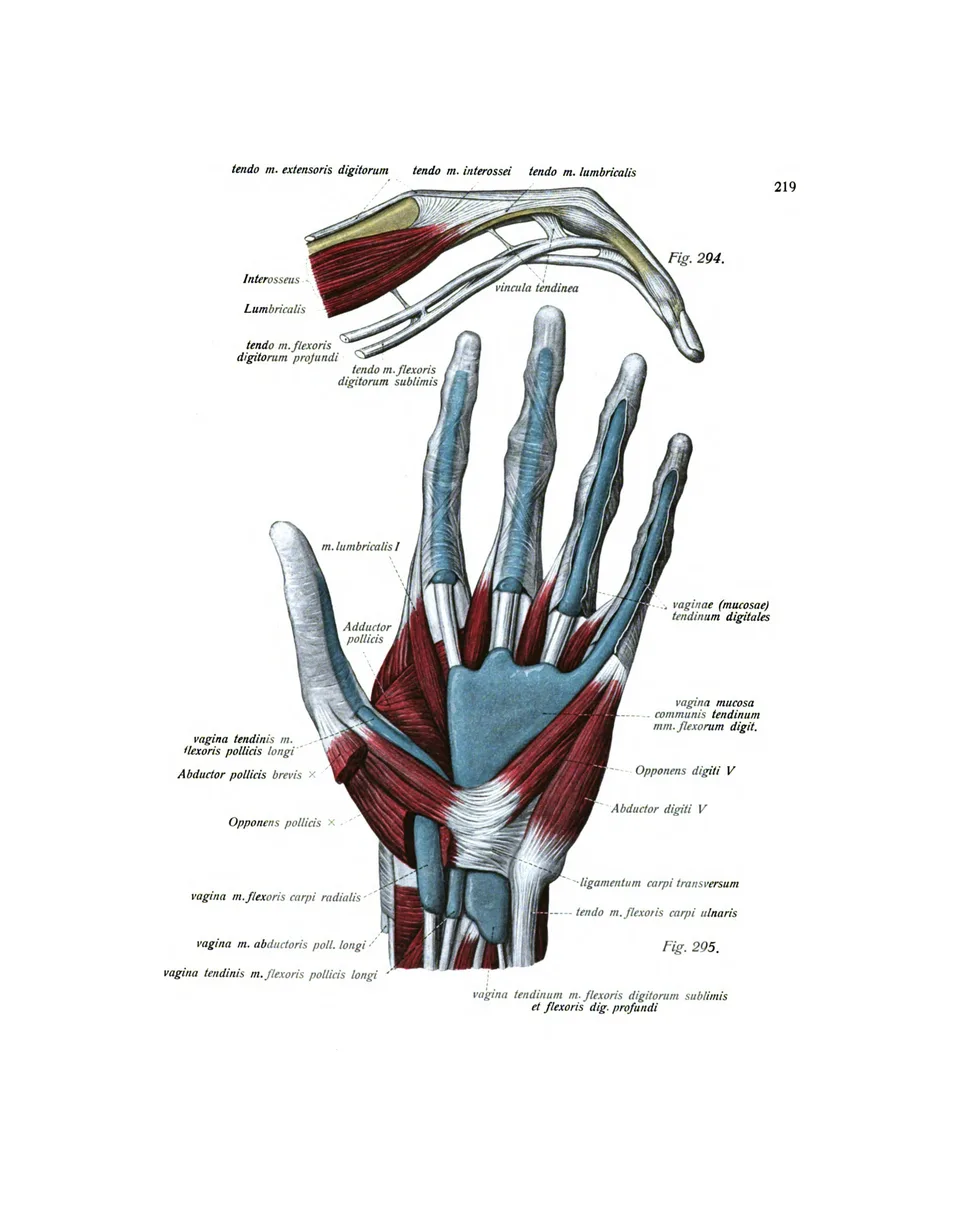
7 plates · Eustachi, Albinus, SobottaThe bones of the hand and wrist, its muscles in layers, and its ligaments and tendons.
The foot
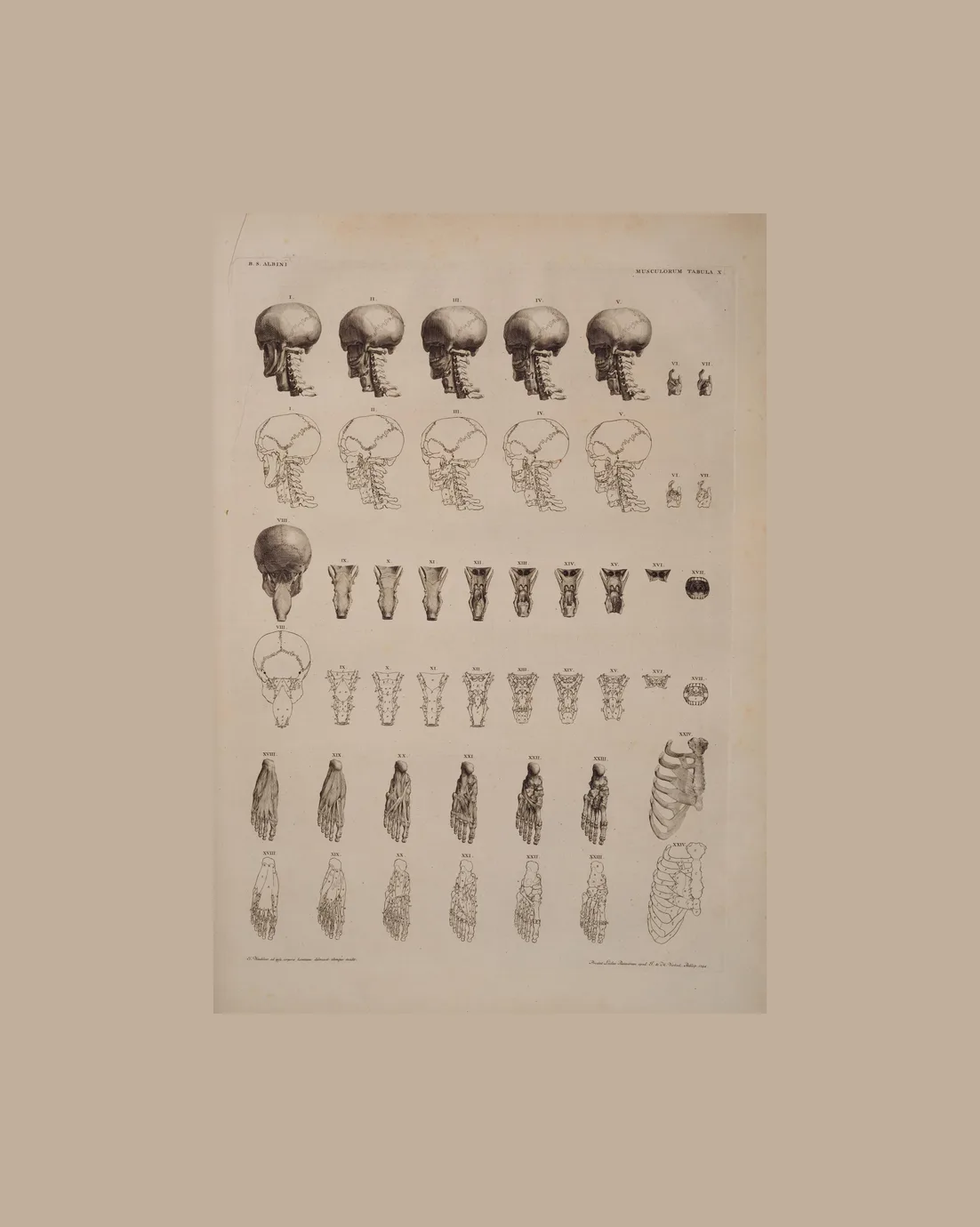
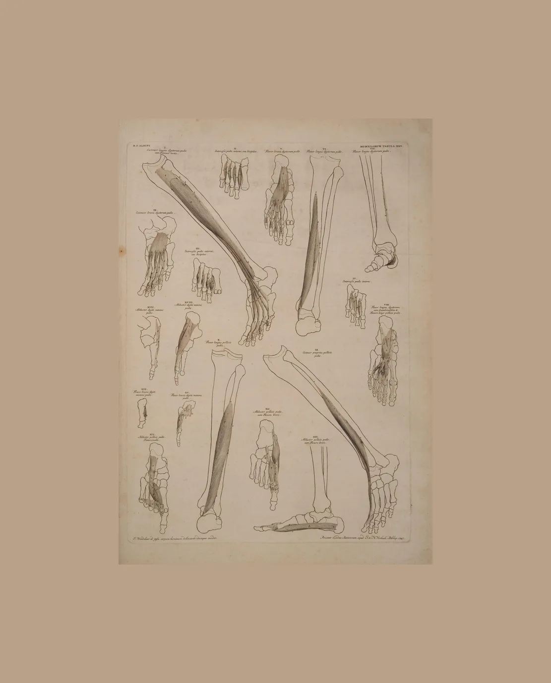
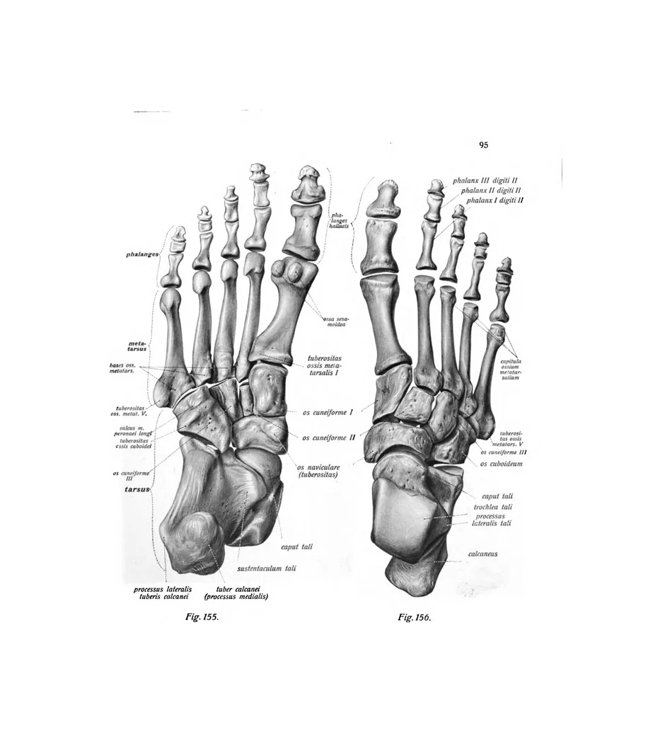
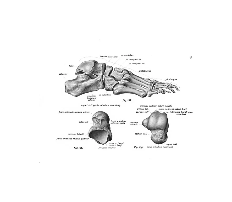
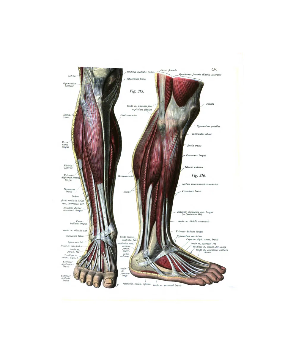
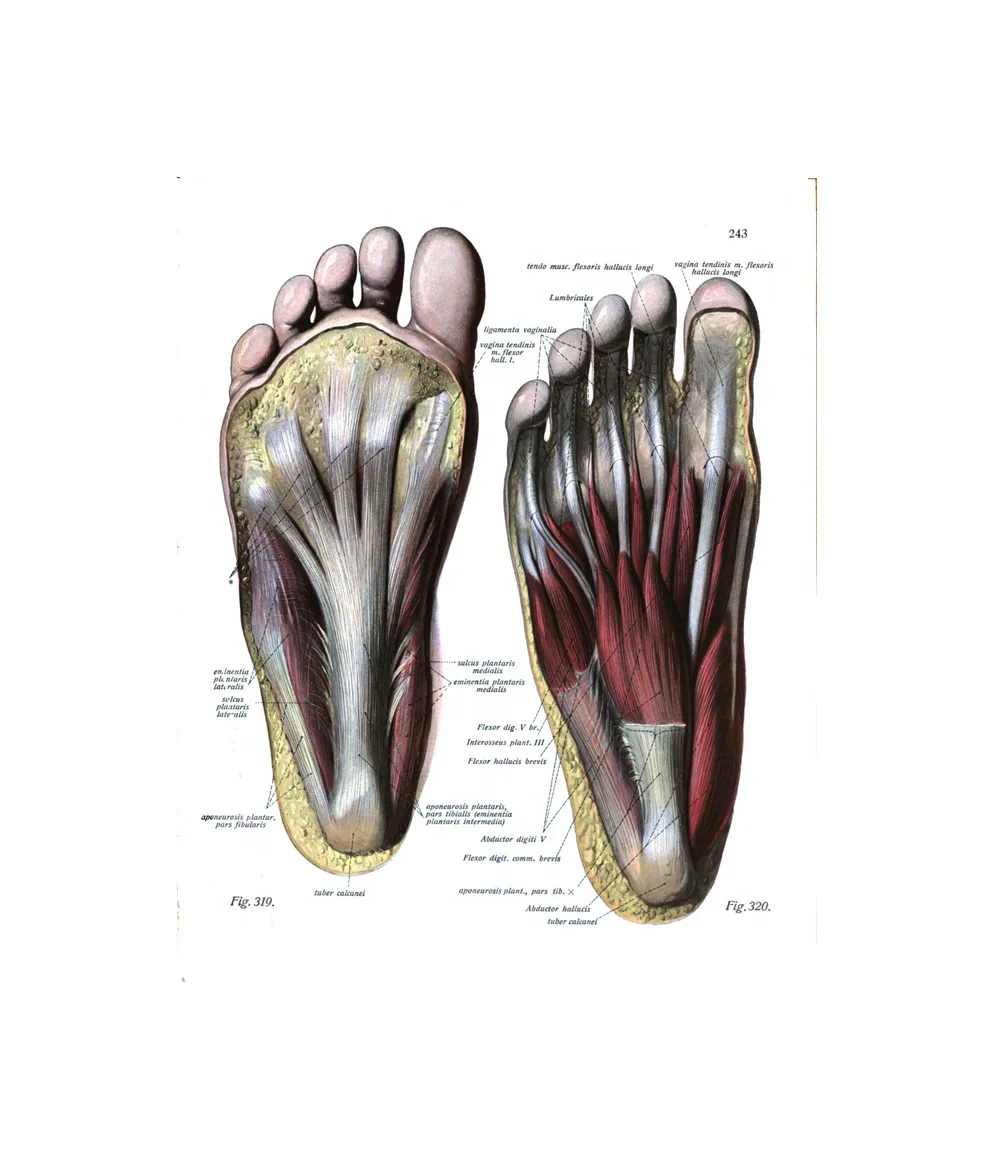
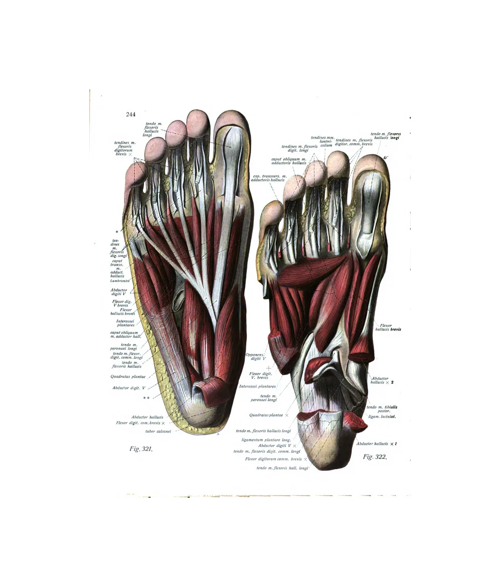
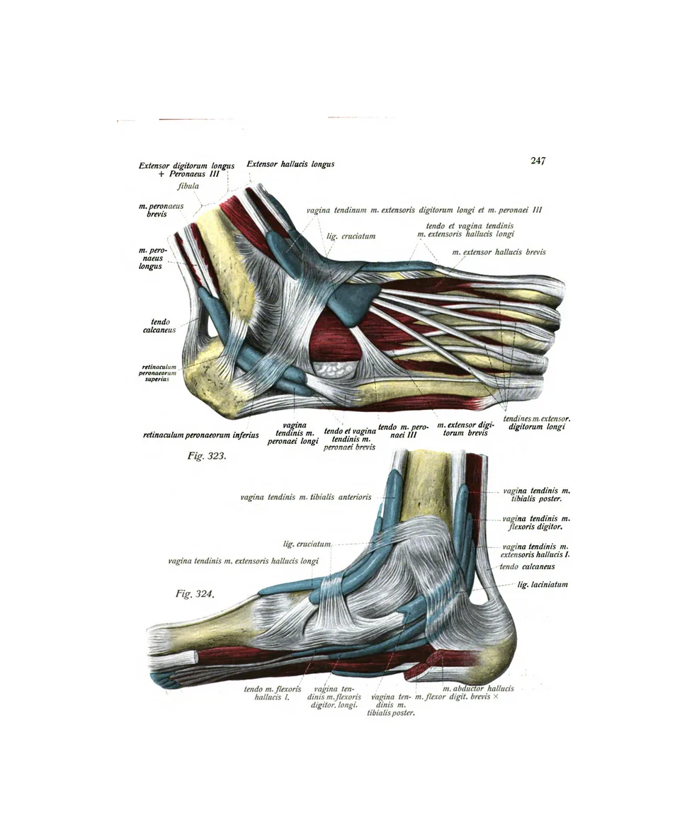
8 plates · Albinus, SobottaThe bones of the foot from above, below and the side, the muscles of the sole and lower leg, and the ligaments of the ankle.
The spine
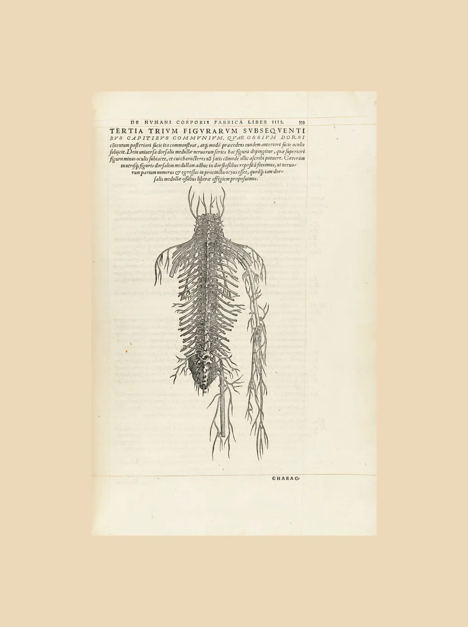
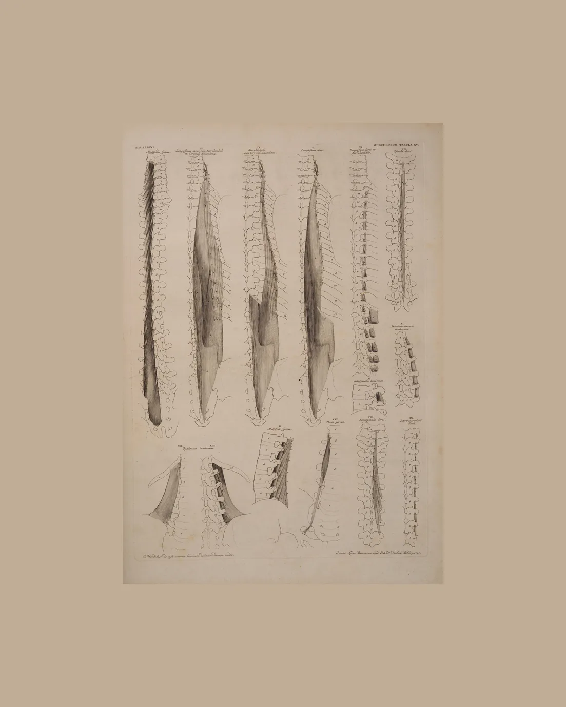
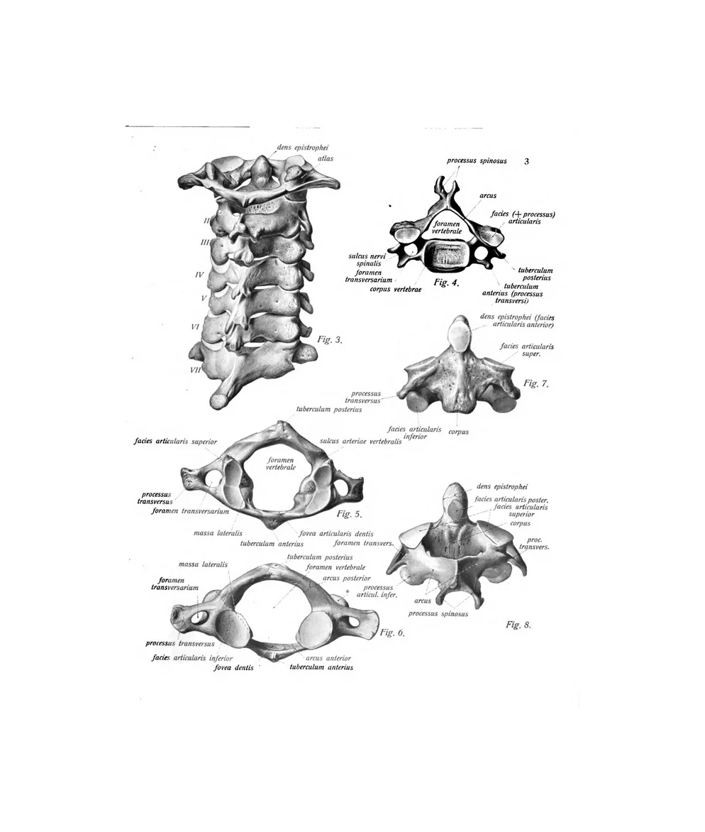
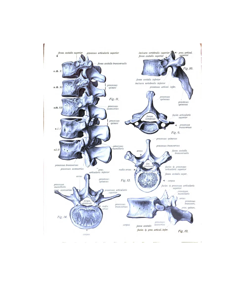
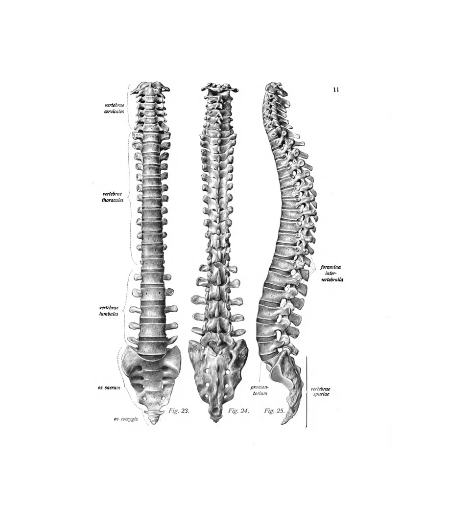
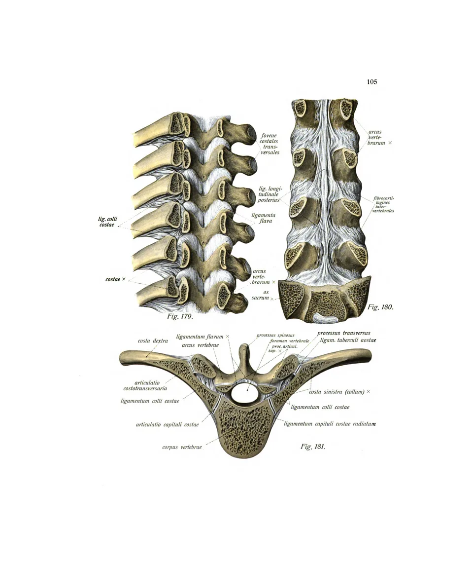
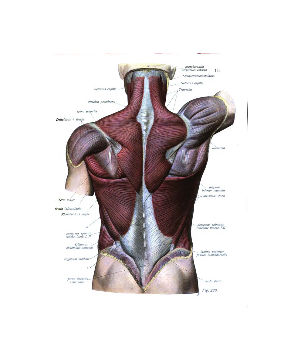
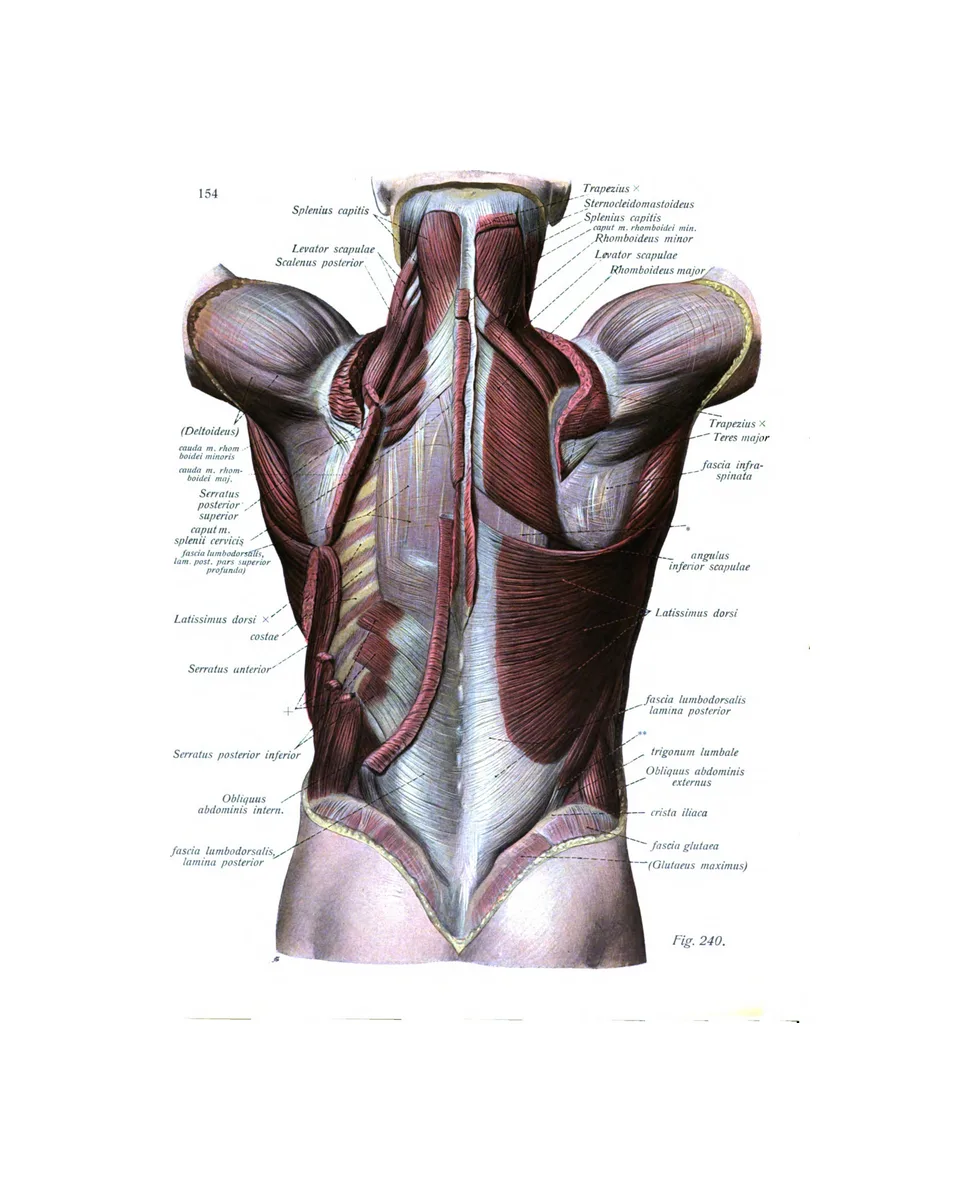
6 plates · Vesalius, Albinus, SobottaThe vertebral column and its vertebrae, its ligaments, the spinal cord and the muscles of the back.
The skeleton and skull
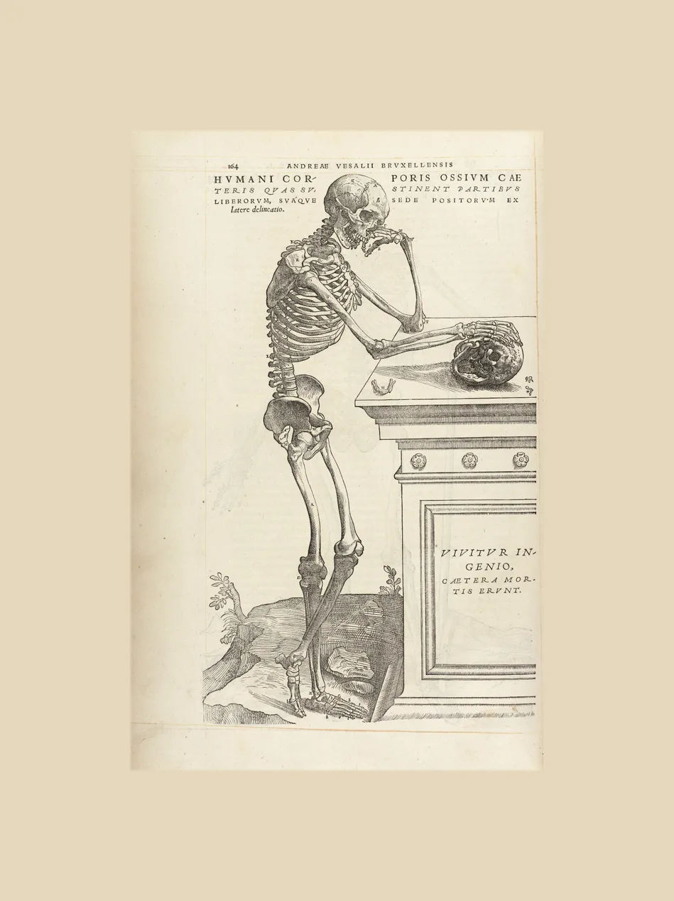
15 plates · Vesalius, Eustachi, Albinus, Parker, SobottaWhole skeletons from all three early atlases, among them Vesalius’s skeleton contemplating a skull and Albinus’s skeletons standing in a landscape.
The organs
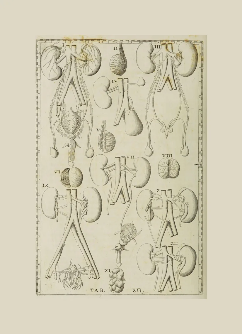
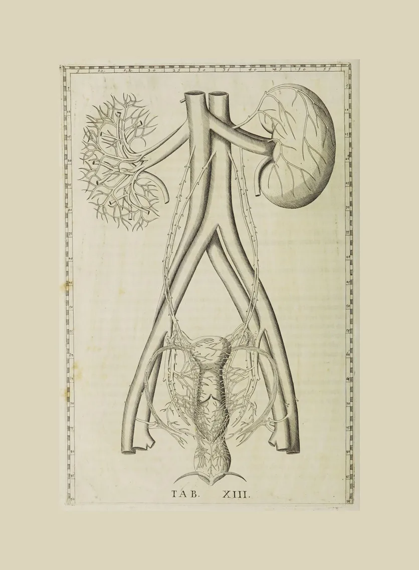
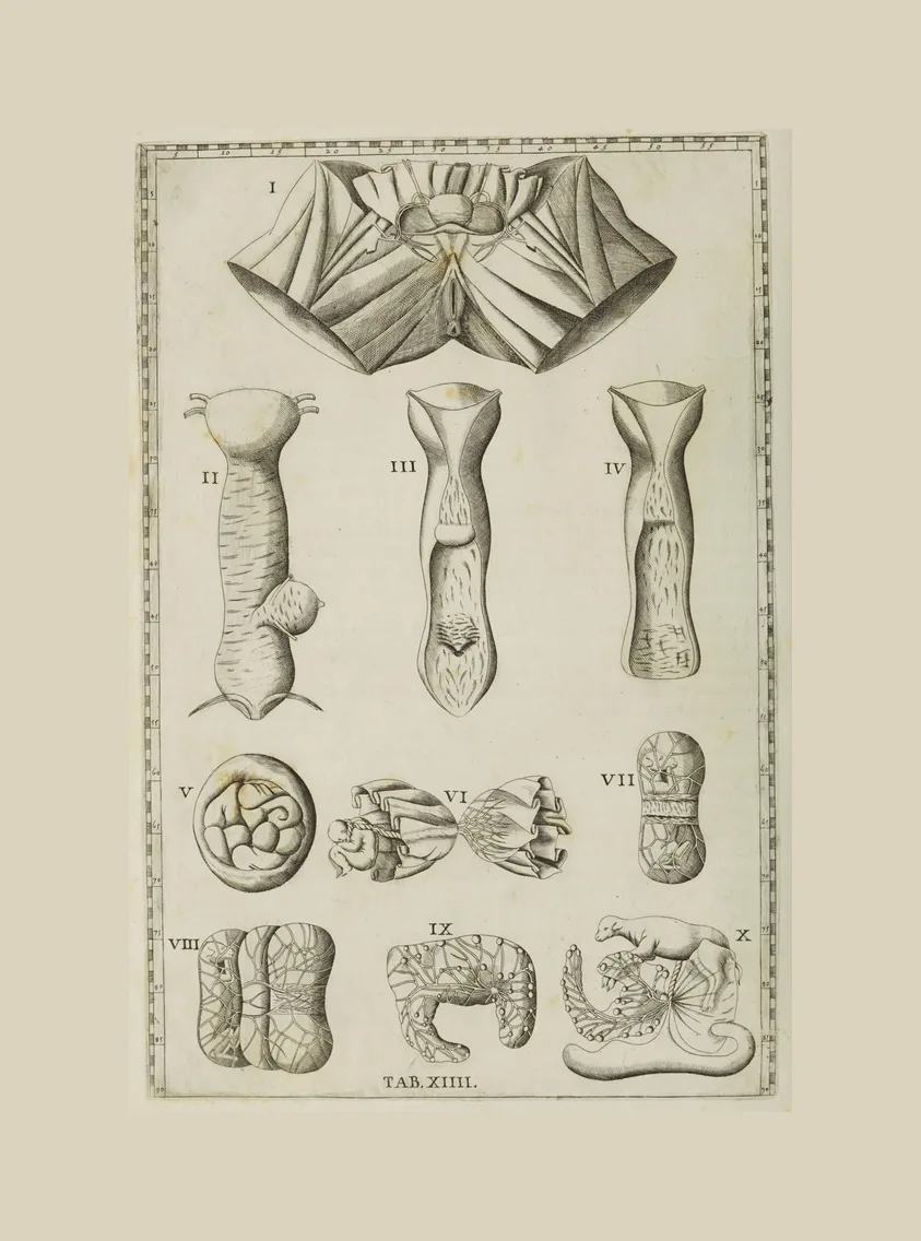
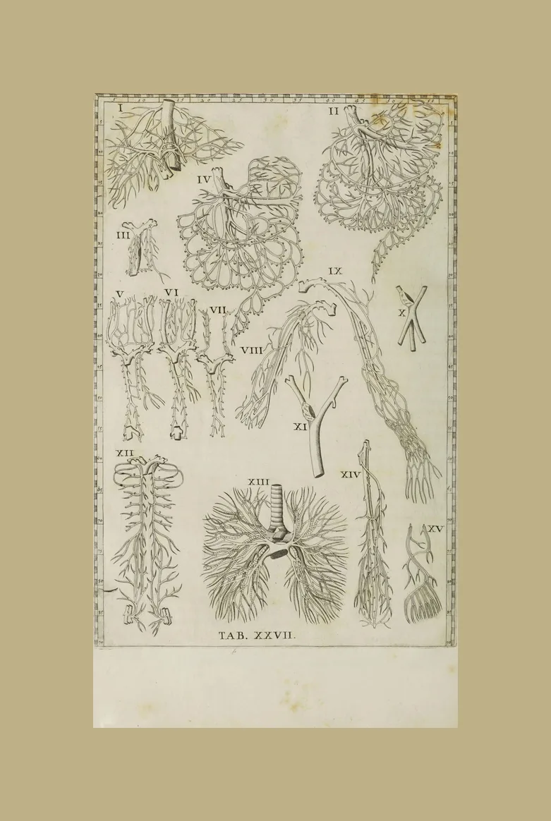
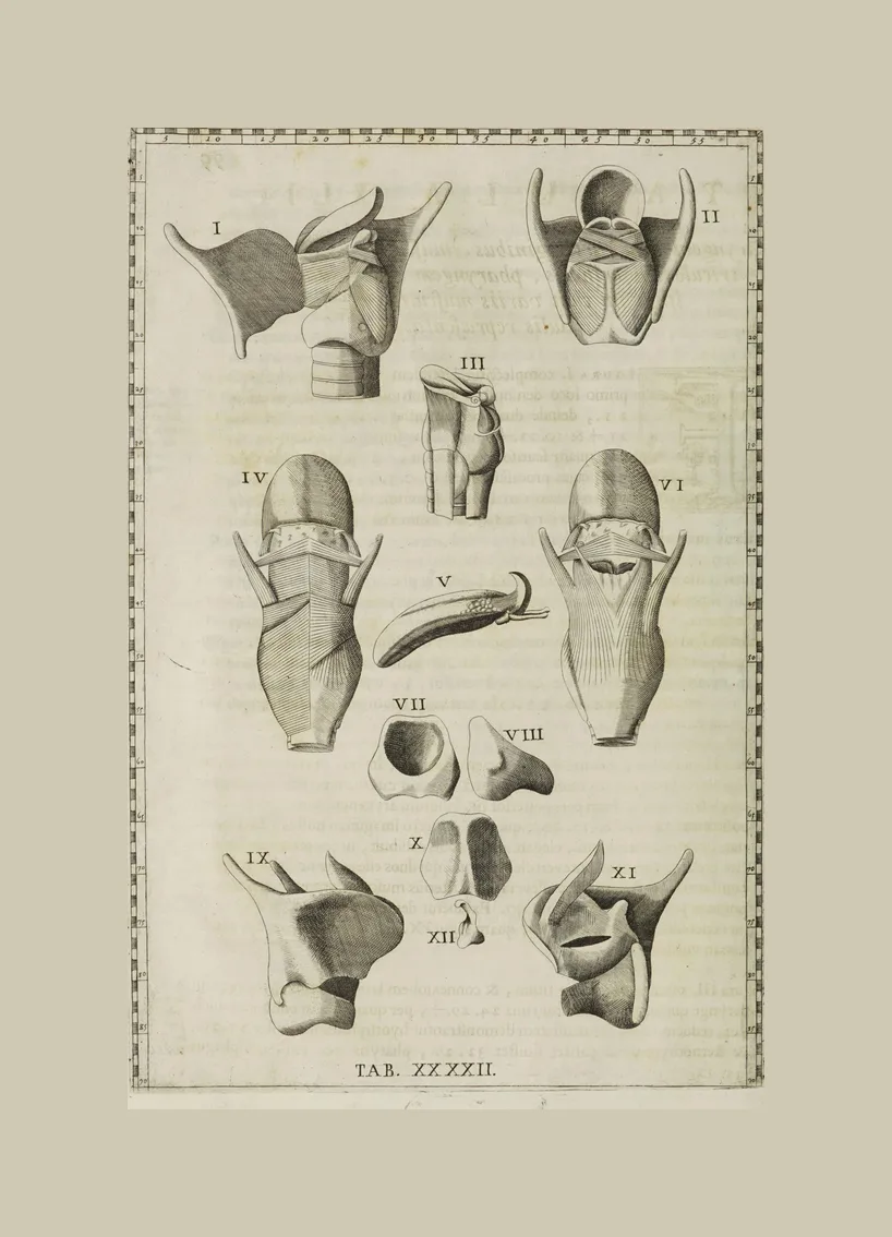
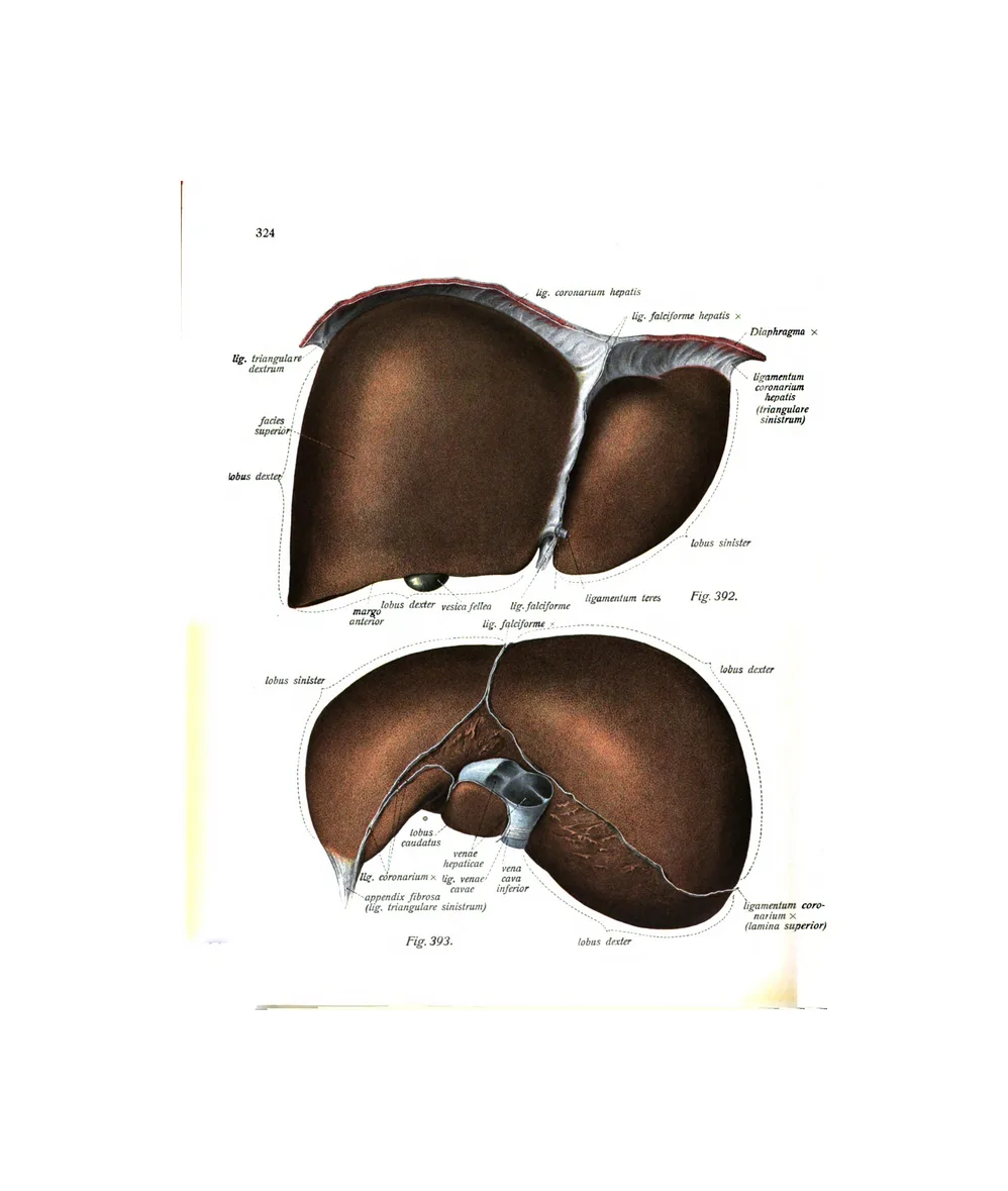
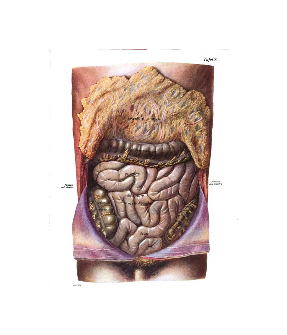
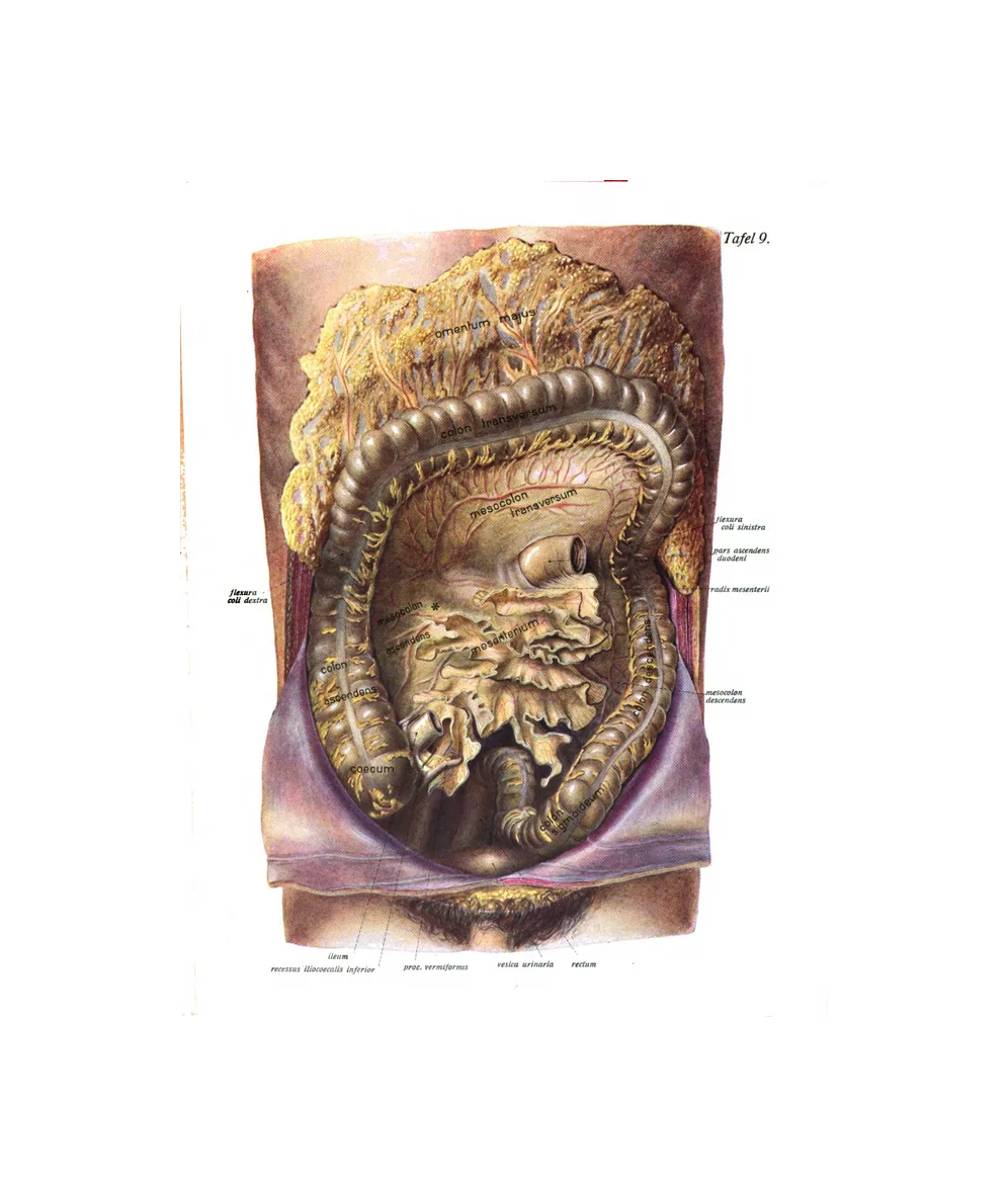
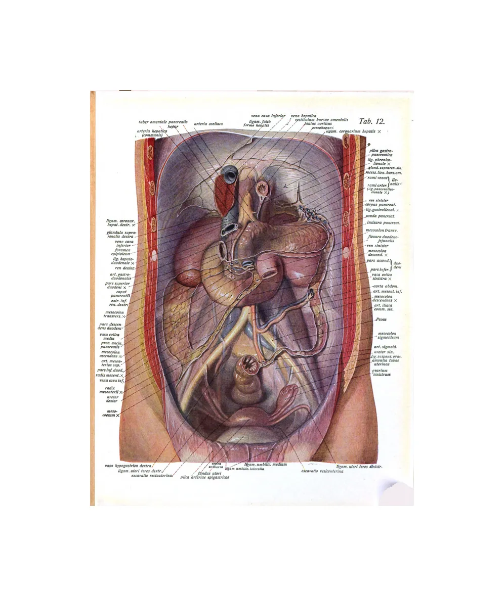
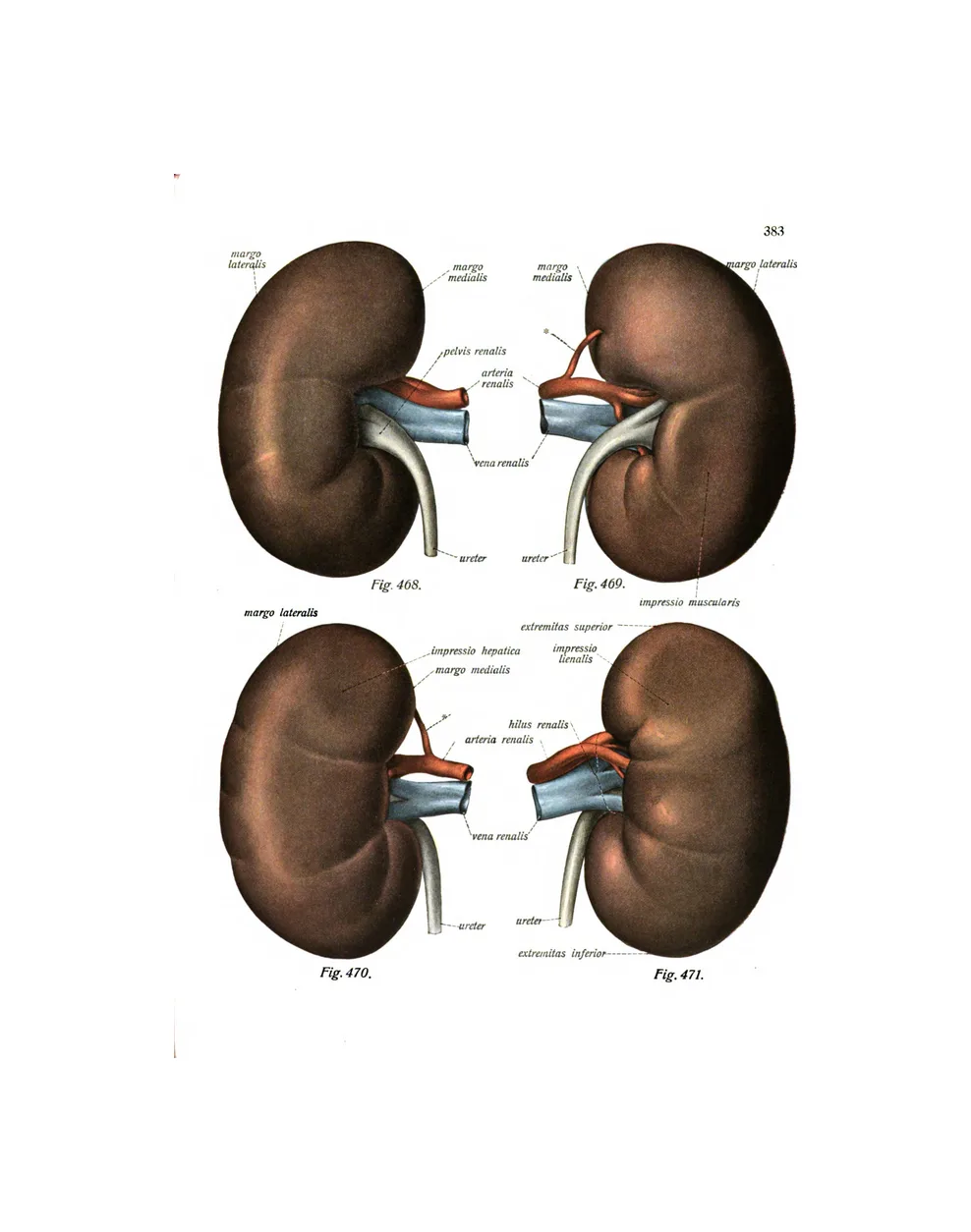
13 plates · Eustachi, SobottaThe stomach, liver, kidneys and intestines, shown in place in the body or removed and drawn on their own.
The muscles
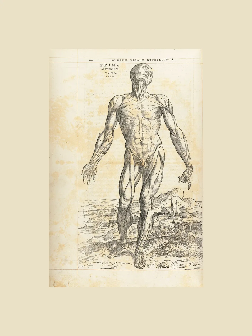
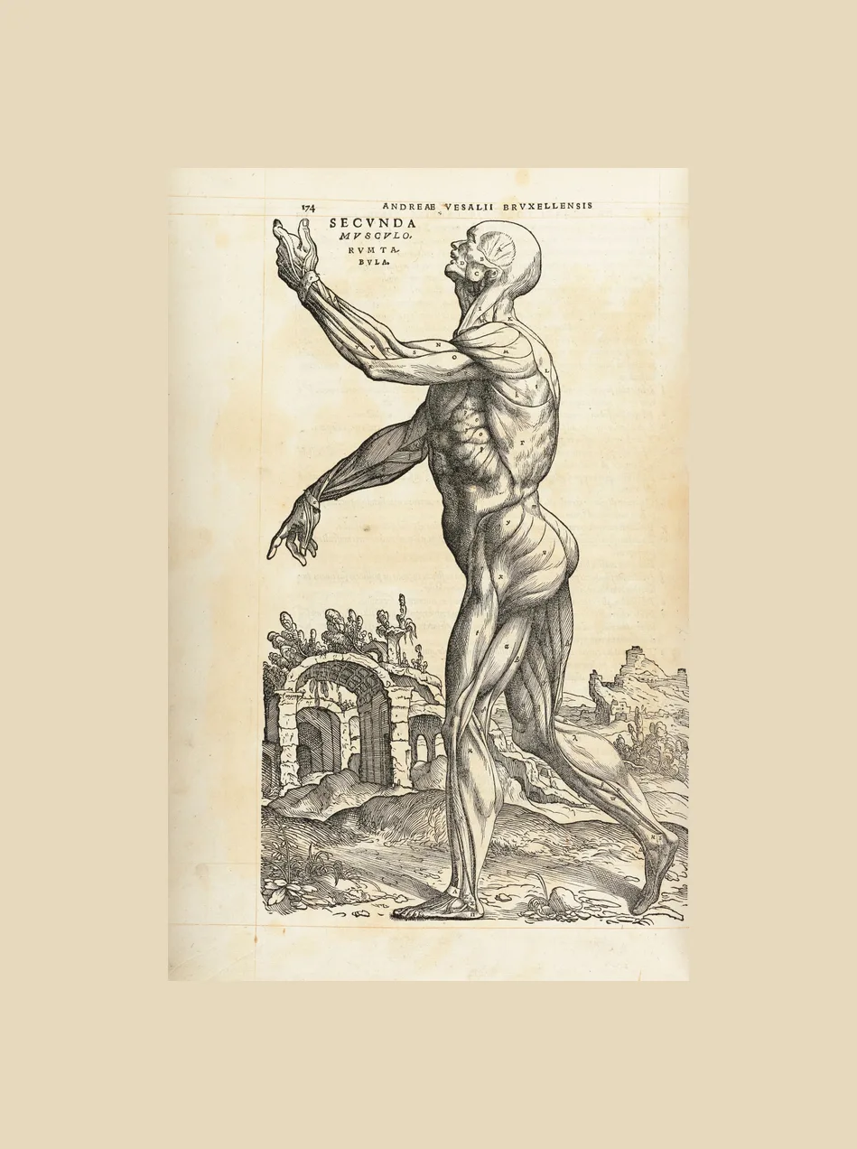
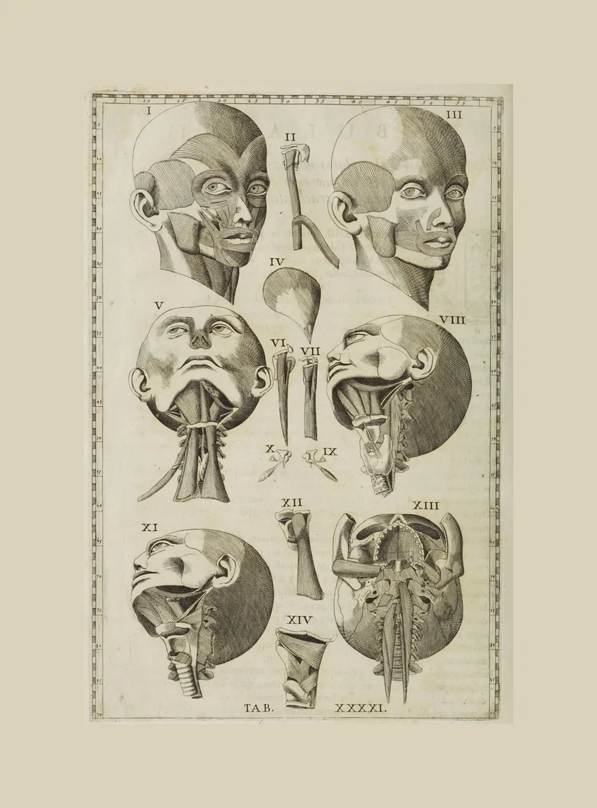
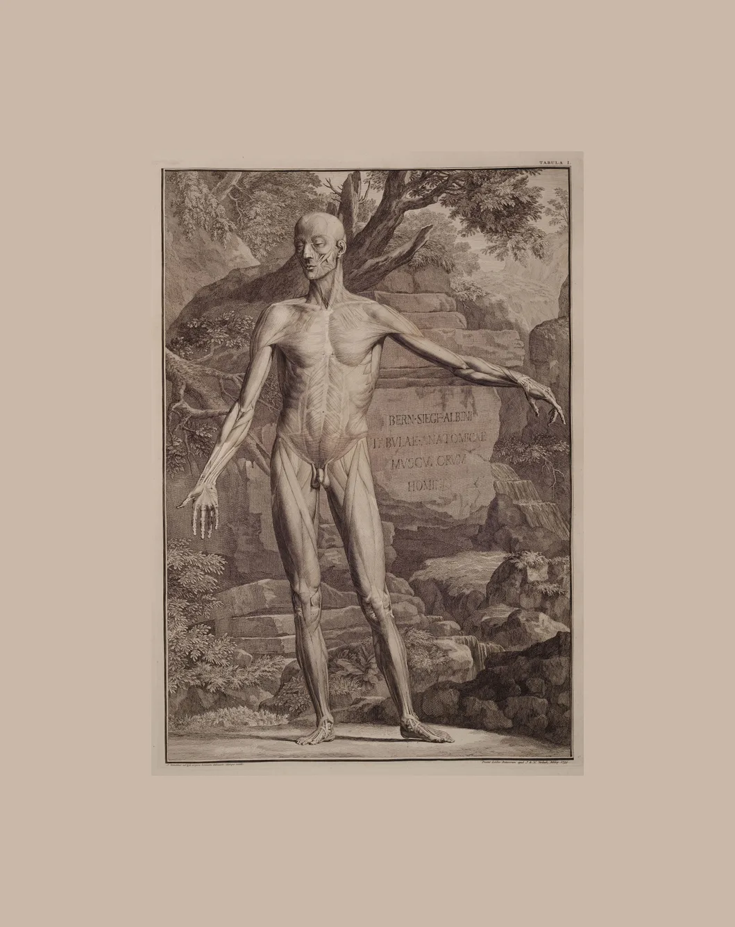
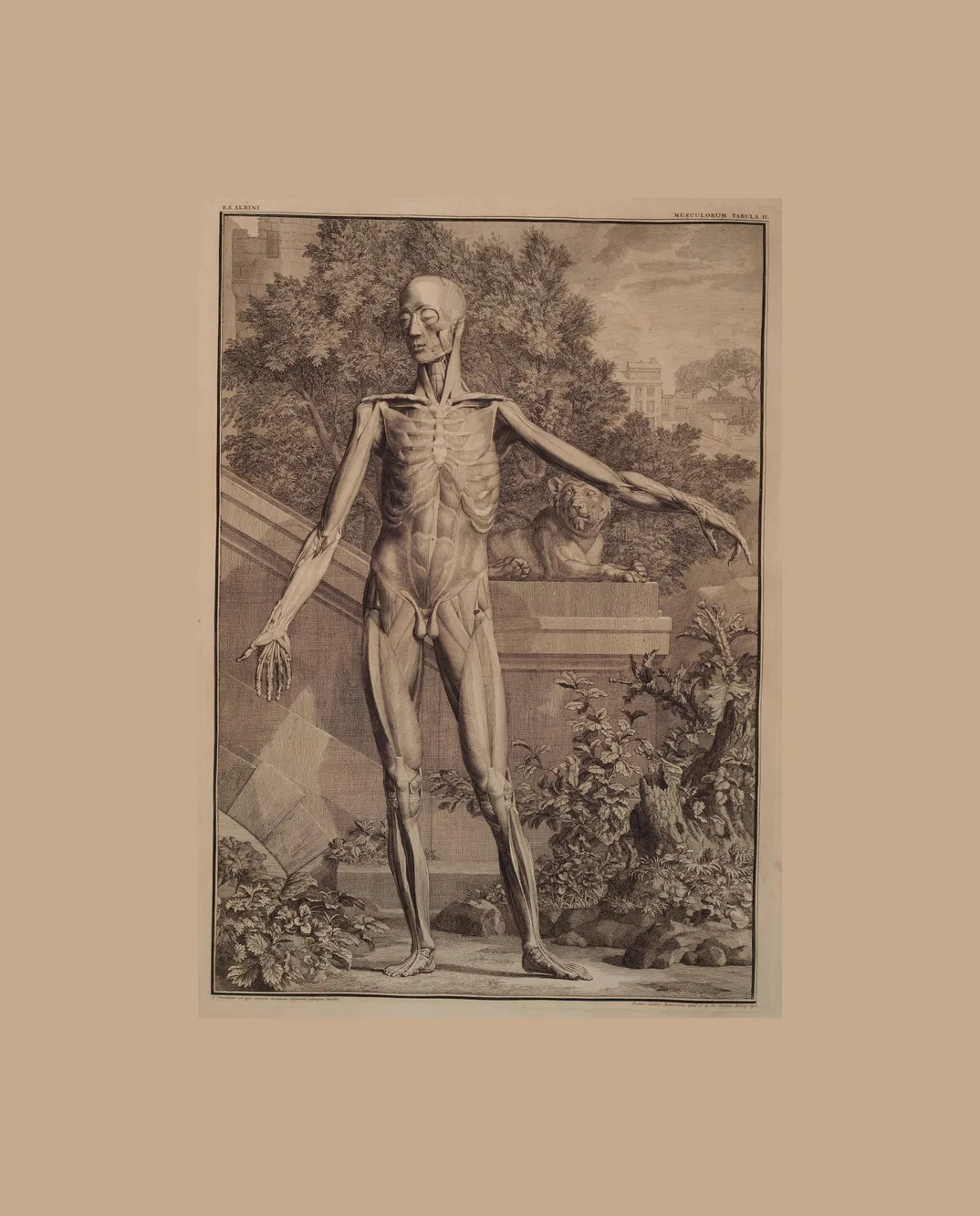
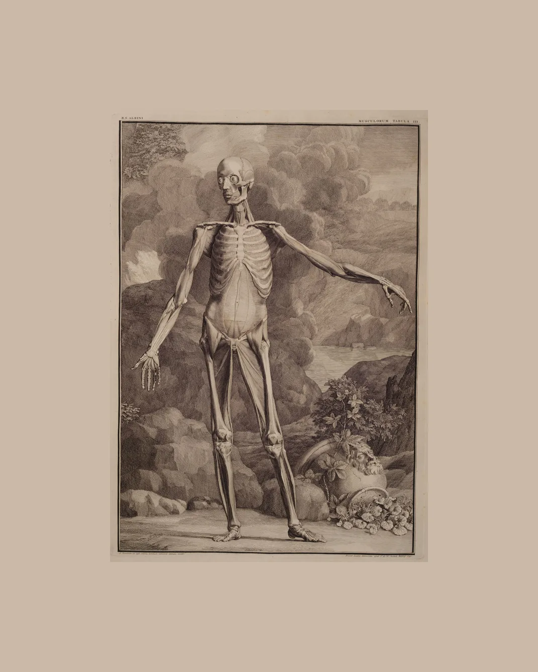
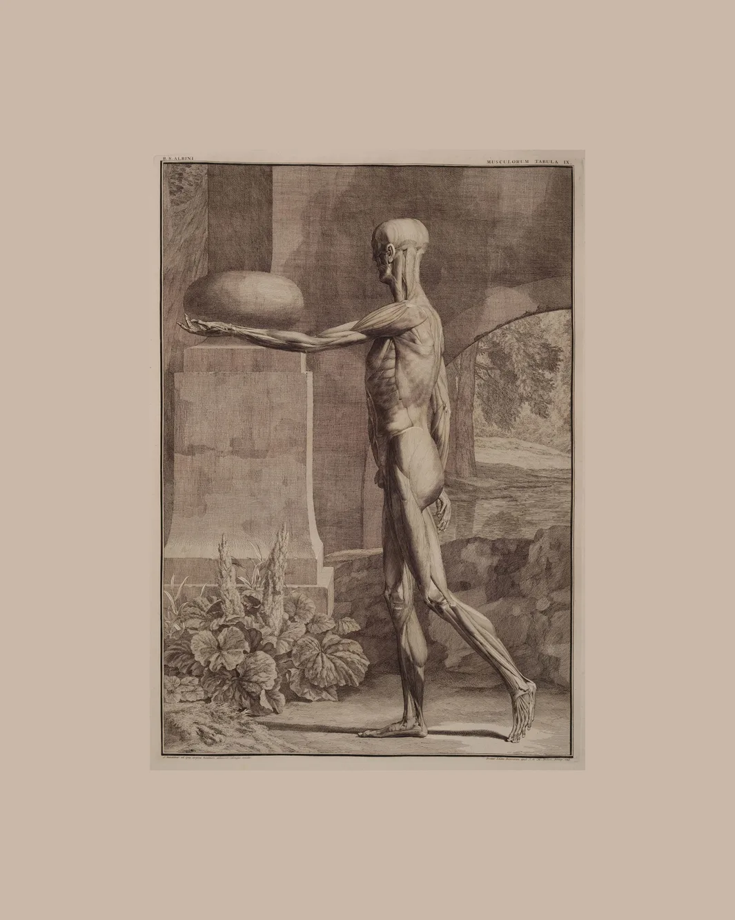
46 plates · Vesalius, Eustachi, Albinus, SobottaThe écorché, the figure drawn with the skin removed, layer by layer. This is the largest group here: Vesalius’s muscle men, Eustachi’s figures in their numbered borders, and Albinus’s four “orders” of muscles.
Vessels and the whole body
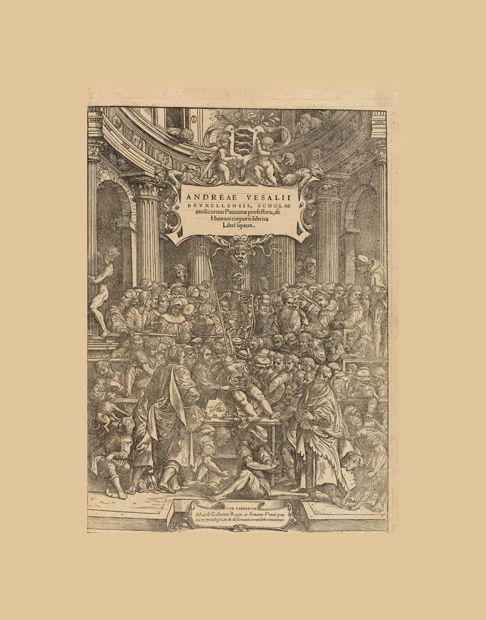
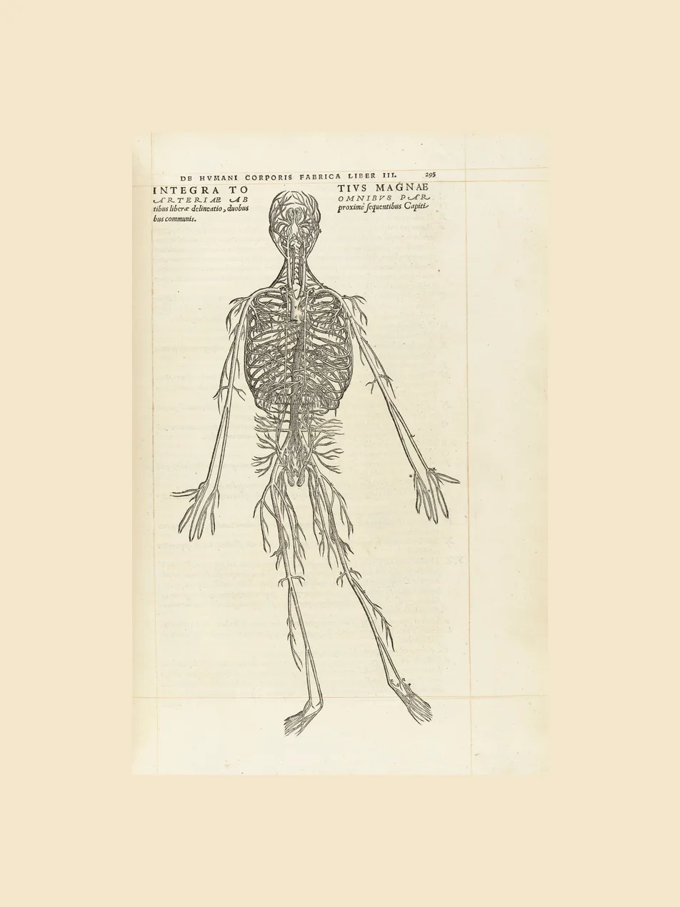
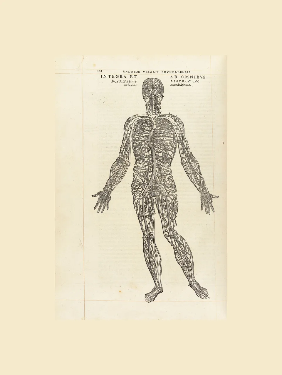
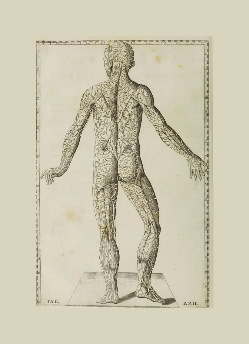
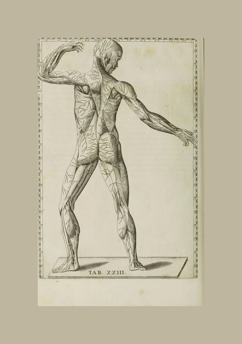
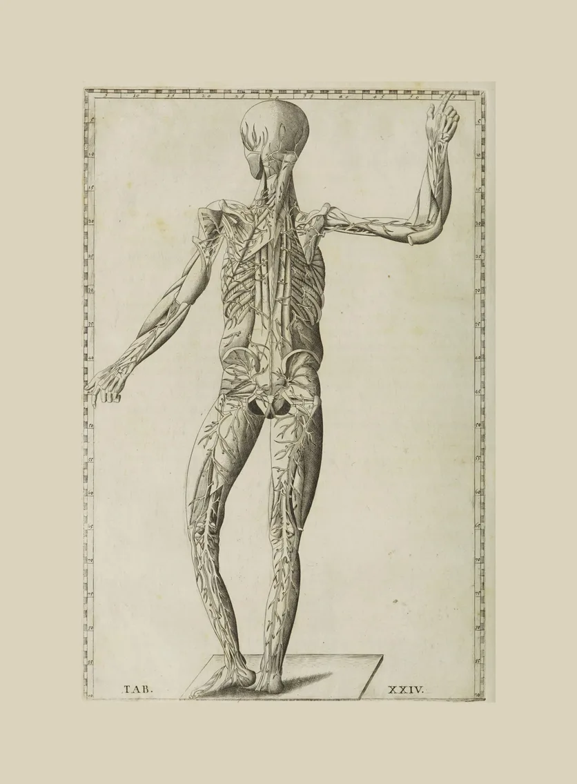
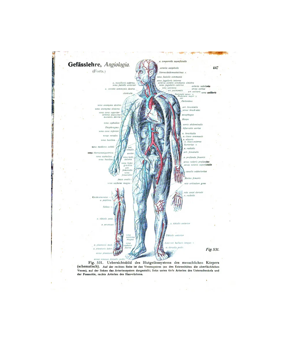
9 plates · Vesalius, Eustachi, SobottaFigures of the whole body that map a single system: the veins, the arteries, and the vessels of the organs. Also here is the title page of the Fabrica, showing a public dissection.
Reading the plate references
Each atlas numbers its pictures in its own way, and the references above follow the original so a plate can be found in a facsimile or a library copy.
- Vesalius’s Fabrica is cited by book and figure. “Book II, third muscle tabula” is the third of the fourteen full-length muscle figures in the second book, which is on the muscles.
- Eustachi is cited by table, Tab. I to XLVII, as Lancisi numbered them in 1714. The numbers in the border of each plate are its coordinate grid, not page numbers.
- Albinus numbers the skeleton tables (Sceleti Tabula) and the muscle tables (Musculorum Tabula) separately.
- Sobotta is cited here by volume and leaf of the scanned 1919–1920 copy at the Internet Archive, which is the easiest way to find the page.
All 151 plates
The full list in one place, for citing. Each title links to its entry above.
| Plate | Atlas | Reference | Year | Body part |
|---|---|---|---|---|
| The Fabrica Title Page (the anatomy theatre) | De humani corporis fabrica | title page | 1543 | Vessels and the whole body |
| The Human Skeleton (front, leaning on a spade) | De humani corporis fabrica | Book I, first skeleton | 1543 | The skeleton and skull |
| The Skeleton Contemplating a Skull | De humani corporis fabrica | Book I, second skeleton | 1543 | The skeleton and skull |
| The First Muscle Man (front) | De humani corporis fabrica | Book II, first muscle tabula | 1543 | The muscles |
| The Second Muscle Man (front, arm raised) | De humani corporis fabrica | Book II, second muscle tabula | 1543 | The muscles |
| The Third Muscle Man (front) | De humani corporis fabrica | Book II, third muscle tabula | 1543 | The muscles |
| The Fourth Muscle Man (front, deeper layer) | De humani corporis fabrica | Book II, fourth muscle tabula | 1543 | The muscles |
| The Fifth Muscle Man (front, deep muscles) | De humani corporis fabrica | Book II, fifth muscle tabula | 1543 | The muscles |
| The Sixth Muscle Man (front, deepest muscles) | De humani corporis fabrica | Book II, sixth muscle tabula | 1543 | The muscles |
| The Seventh Muscle Man (hanging figure) | De humani corporis fabrica | Book II, seventh muscle tabula | 1543 | The muscles |
| The Eighth Muscle Man (suspended by the wall) | De humani corporis fabrica | Book II, eighth muscle tabula | 1543 | The muscles |
| The Ninth Muscle Man (back) | De humani corporis fabrica | Book II, ninth muscle tabula | 1543 | The muscles |
| The Tenth Muscle Man (back, arm raised) | De humani corporis fabrica | Book II, tenth muscle tabula | 1543 | The muscles |
| The Eleventh Muscle Man (back, deeper layer) | De humani corporis fabrica | Book II, eleventh muscle tabula | 1543 | The muscles |
| The Twelfth Muscle Man (back, deep muscles) | De humani corporis fabrica | Book II, twelfth muscle tabula | 1543 | The muscles |
| The Thirteenth Muscle Man (back, deepest muscles) | De humani corporis fabrica | Book II, thirteenth muscle tabula | 1543 | The muscles |
| The Fourteenth Muscle Man (back, bowed) | De humani corporis fabrica | Book II, fourteenth muscle tabula | 1543 | The muscles |
| The Arterial System (the artery man) | De humani corporis fabrica | Book III, artery figure | 1543 | Vessels and the whole body |
| The Venous System (the vein man) | De humani corporis fabrica | Book III, vein figure | 1543 | Vessels and the whole body |
| The Nervous System (the thirty pairs of nerves) | De humani corporis fabrica | Book IV, nerve figure | 1543 | The brain and nerves |
| The Spinal Cord and Spinal Nerves | De humani corporis fabrica | Book IV, third nerve figure | 1543 | The spine |
| Figure of a Man with the Viscera in Place | Tabulae anatomicae | Tab. IX | 1714 | The organs |
| The Stomach and Intestines | Tabulae anatomicae | Tab. X | 1714 | The organs |
| The Liver, Spleen and Stomach | Tabulae anatomicae | Tab. XI | 1714 | The organs |
| The Kidneys, Bladder and Their Vessels | Tabulae anatomicae | Tab. XII | 1714 | The organs |
| The Kidneys with the Aorta and Vena Cava | Tabulae anatomicae | Tab. XIII | 1714 | The organs |
| The Diaphragm, Oesophagus and Foetal Membranes (Tab. XIV) | Tabulae anatomicae | Tab. XIV | 1714 | The organs |
| The Lungs, Trachea and Heart | Tabulae anatomicae | Tab. XV | 1714 | The lungs and chest |
| The Human Heart (six anatomical views) | Tabulae anatomicae | Tab. XVI | 1714 | The heart |
| The Brain in Section (seven views) | Tabulae anatomicae | Tab. XVII | 1714 | The brain and nerves |
| The Nervous System (brain, spinal cord & nerves) | Tabulae anatomicae | Tab. XVIII | 1714 | The brain and nerves |
| The Spinal Nerves (figure from behind) | Tabulae anatomicae | Tab. XIX | 1714 | The brain and nerves |
| The Muscular System (posterior écorché) | Tabulae anatomicae | Tab. XX | 1714 | The muscles |
| The Muscular System (dynamic écorché) | Tabulae anatomicae | Tab. XXI | 1714 | The muscles |
| Veins of the Body, Seen from Behind (Tab. XXII) | Tabulae anatomicae | Tab. XXII | 1714 | Vessels and the whole body |
| Veins of the Body (figure from the side) | Tabulae anatomicae | Tab. XXIII | 1714 | Vessels and the whole body |
| Veins of the Body (figure from behind) | Tabulae anatomicae | Tab. XXIV | 1714 | Vessels and the whole body |
| Arteries of the Body (figure with arms raised) | Tabulae anatomicae | Tab. XXV | 1714 | Vessels and the whole body |
| Arteries of the Body (figure from the front) | Tabulae anatomicae | Tab. XXVI | 1714 | Vessels and the whole body |
| Blood Vessels of the Viscera (fifteen dissections) | Tabulae anatomicae | Tab. XXVII | 1714 | The organs |
| The Muscular System (anterior muscle figure) | Tabulae anatomicae | Tab. XXVIII | 1714 | The muscles |
| Écorché Muscle Man (posterior view) | Tabulae anatomicae | Tab. XXIX | 1714 | The muscles |
| Écorché Muscle Man (anterior, alternate pose) | Tabulae anatomicae | Tab. XXX | 1714 | The muscles |
| Écorché Muscle Man (turning, from behind) | Tabulae anatomicae | Tab. XXXI | 1714 | The muscles |
| Muscles of the Body, Second Layer (front) | Tabulae anatomicae | Tab. XXXII | 1714 | The muscles |
| Deep Muscles with the Skeleton (arm extended) | Tabulae anatomicae | Tab. XXXIII | 1714 | The muscles |
| Deep Muscles and Skeleton, from Behind (Tab. XXXIV) | Tabulae anatomicae | Tab. XXXIV | 1714 | The muscles |
| Deepest Muscles of the Body (front, on the skeleton) | Tabulae anatomicae | Tab. XXXV | 1714 | The muscles |
| Deep Muscles of the Body (back) | Tabulae anatomicae | Tab. XXXVI | 1714 | The muscles |
| Deep Muscles of the Back (arm raised) | Tabulae anatomicae | Tab. XXXVII | 1714 | The muscles |
| Deep Muscles and Skeleton, from the Front, Head Raised (Tab. XXXVIII) | Tabulae anatomicae | Tab. XXXVIII | 1714 | The muscles |
| Deep Muscles of the Back (with skeleton) | Tabulae anatomicae | Tab. XXXIX | 1714 | The muscles |
| The Human Eye (twelve dissections) | Tabulae anatomicae | Tab. XL | 1714 | The eye and ear |
| Muscles of the Face, Eye and Throat | Tabulae anatomicae | Tab. XLI | 1714 | The muscles |
| The Larynx and Hyoid (fourteen dissections) | Tabulae anatomicae | Tab. XLII | 1714 | The organs |
| The Human Skeleton (front view) | Tabulae anatomicae | Tab. XLIII | 1714 | The skeleton and skull |
| The Human Skeleton (back view) | Tabulae anatomicae | Tab. XLIV | 1714 | The skeleton and skull |
| The Human Skeleton (side view, walking) | Tabulae anatomicae | Tab. XLV | 1714 | The skeleton and skull |
| The Human Skull (seventeen views) | Tabulae anatomicae | Tab. XLVI | 1714 | The teeth and jaw |
| Bones of the Body & the Hand (skeletal plate) | Tabulae anatomicae | Tab. XLVII | 1714 | The hand |
| The Human Skeleton (front view, in a landscape) | Tabulae sceleti et musculorum corporis humani | Sceleti Tabula I | 1747 | The skeleton and skull |
| The Human Skeleton (back view, in a landscape) | Tabulae sceleti et musculorum corporis humani | Sceleti Tabula II | 1747 | The skeleton and skull |
| The Human Skeleton (side view, in a landscape) | Tabulae sceleti et musculorum corporis humani | Sceleti Tabula III | 1747 | The skeleton and skull |
| Muscles of the Body, First Order (front) | Tabulae sceleti et musculorum corporis humani | Musculorum Tabula I | 1747 | The muscles |
| Muscles of the Body, Second Order (front) | Tabulae sceleti et musculorum corporis humani | Musculorum Tabula II | 1747 | The muscles |
| Muscles of the Body, Third Order (front) | Tabulae sceleti et musculorum corporis humani | Musculorum Tabula III | 1747 | The muscles |
| Fourth Order of Muscles, Front, with the Rhinoceros | Tabulae sceleti et musculorum corporis humani | Musculorum Tabula IV | 1747 | The muscles |
| Muscles of the Body, First Order (back) | Tabulae sceleti et musculorum corporis humani | Musculorum Tabula V | 1747 | The muscles |
| Muscles of the Body, Second Order (back) | Tabulae sceleti et musculorum corporis humani | Musculorum Tabula VI | 1747 | The muscles |
| Muscles of the Body, Third Order (back) | Tabulae sceleti et musculorum corporis humani | Musculorum Tabula VII | 1747 | The muscles |
| Fourth Order of Muscles, Back, with the Rhinoceros | Tabulae sceleti et musculorum corporis humani | Musculorum Tabula VIII | 1747 | The muscles |
| Muscles of the Body (side view, arm extended) | Tabulae sceleti et musculorum corporis humani | Musculorum Tabula IX | 1747 | The muscles |
| Muscles of the Neck, Larynx and Foot (study plate) | Tabulae sceleti et musculorum corporis humani | Musculorum Tabula X | 1747 | The foot |
| Muscles of the Face, Ear and Jaw (study plate) | Tabulae sceleti et musculorum corporis humani | Musculorum Tabula XI | 1747 | The eye and ear |
| Muscles of the Larynx, Jaw and Pharynx (study plate) | Tabulae sceleti et musculorum corporis humani | Musculorum Tabula XII | 1747 | The teeth and jaw |
| Muscles of the Spine and Back | Tabulae sceleti et musculorum corporis humani | Musculorum Tabula XV | 1747 | The spine |
| Muscles of the Hand and Forearm | Tabulae sceleti et musculorum corporis humani | Musculorum Tabula XX | 1747 | The hand |
| Muscles of the Foot and Lower Leg | Tabulae sceleti et musculorum corporis humani | Musculorum Tabula XXV | 1747 | The foot |
| Anatomical Plates: Skeleton & Arteries (front and back) | Anatomical Plates | single sheet | 1830 | The skeleton and skull |
| The Cervical Vertebrae (atlas and axis) | Atlas der deskriptiven Anatomie des Menschen | vol. I, leaf 14 | 1919 | The spine |
| Vertebrae of the Spine (thoracic and lumbar) | Atlas der deskriptiven Anatomie des Menschen | vol. I, leaf 15 | 1919 | The spine |
| The Vertebral Column (three views) | Atlas der deskriptiven Anatomie des Menschen | vol. I, leaf 22 | 1919 | The spine |
| The Thorax: Ribs, Sternum and Spine | Atlas der deskriptiven Anatomie des Menschen | vol. I, leaf 27 | 1919 | The skeleton and skull |
| The Skull (front view) | Atlas der deskriptiven Anatomie des Menschen | vol. I, leaf 31 | 1919 | The skeleton and skull |
| The Skull (side view) | Atlas der deskriptiven Anatomie des Menschen | vol. I, leaf 32 | 1919 | The skeleton and skull |
| The Base of the Skull (from below) | Atlas der deskriptiven Anatomie des Menschen | vol. I, leaf 33 | 1919 | The skeleton and skull |
| The Base of the Skull (interior) | Atlas der deskriptiven Anatomie des Menschen | vol. I, leaf 34 | 1919 | The skeleton and skull |
| Bones of the Hand and Wrist (separated) | Atlas der deskriptiven Anatomie des Menschen | vol. I, leaf 64 | 1919 | The hand |
| Bones of the Hand (palm and back) | Atlas der deskriptiven Anatomie des Menschen | vol. I, leaf 65 | 1919 | The hand |
| Bones of the Foot (from above and below) | Atlas der deskriptiven Anatomie des Menschen | vol. I, leaf 80 | 1919 | The foot |
| Bones of the Foot (side view, with the talus and heel bone) | Atlas der deskriptiven Anatomie des Menschen | vol. I, leaf 81 | 1919 | The foot |
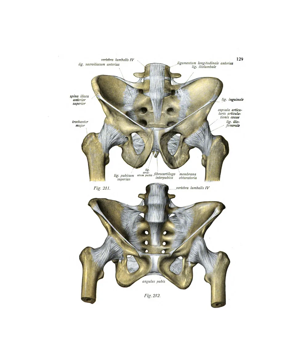
| Ligaments of the Spine (vertebral joints) | Atlas der deskriptiven Anatomie des Menschen | vol. I, leaf 90 | 1919 | The spine |
| The Pelvis (bones of the hip) | Atlas der deskriptiven Anatomie des Menschen | vol. I, leaf 114 | 1919 | The skeleton and skull |
| Muscles of the Back, Superficial Layer | Atlas der deskriptiven Anatomie des Menschen | vol. I, leaf 138 | 1919 | The muscles |
| Muscles of the Back, Second Layer | Atlas der deskriptiven Anatomie des Menschen | vol. I, leaf 139 | 1919 | The muscles |
| Muscles of the Back, Deep Layer | Atlas der deskriptiven Anatomie des Menschen | vol. I, leaf 142 | 1919 | The muscles |
| Muscles of the Chest and Shoulder | Atlas der deskriptiven Anatomie des Menschen | vol. I, leaf 150 | 1919 | The muscles |
| Muscles of the Head and Neck (side view) | Atlas der deskriptiven Anatomie des Menschen | vol. I, leaf 166 | 1919 | The muscles |
| Deep Muscles of the Neck and Face | Atlas der deskriptiven Anatomie des Menschen | vol. I, leaf 167 | 1919 | The muscles |
| Muscles of the Forearm and Elbow (front, two layers) | Atlas der deskriptiven Anatomie des Menschen | vol. I, leaf 185 | 1919 | The muscles |
| Muscles of the Hand (superficial layer) | Atlas der deskriptiven Anatomie des Menschen | vol. I, leaf 193 | 1919 | The hand |
| Muscles of the Hand (deep layers) | Atlas der deskriptiven Anatomie des Menschen | vol. I, leaf 196 | 1919 | The hand |
| Ligaments and Tendons of the Hand | Atlas der deskriptiven Anatomie des Menschen | vol. I, leaf 200 | 1919 | The hand |
| Muscles of the Thigh (two front views) | Atlas der deskriptiven Anatomie des Menschen | vol. I, leaf 211 | 1919 | The muscles |
| Muscles of the Lower Leg and Foot (side view) | Atlas der deskriptiven Anatomie des Menschen | vol. I, leaf 222 | 1919 | The foot |
| Muscles of the Sole of the Foot | Atlas der deskriptiven Anatomie des Menschen | vol. I, leaf 226 | 1919 | The foot |
| Deep Muscles of the Sole of the Foot | Atlas der deskriptiven Anatomie des Menschen | vol. I, leaf 227 | 1919 | The foot |
| Ligaments of the Foot and Ankle | Atlas der deskriptiven Anatomie des Menschen | vol. I, leaf 230 | 1919 | The foot |
| The Permanent Teeth (upper and lower dentition) | Atlas der deskriptiven Anatomie des Menschen | vol. II, leaf 22 | 1920 | The teeth and jaw |
| The Milk Teeth and Permanent Teeth | Atlas der deskriptiven Anatomie des Menschen | vol. II, leaf 23 | 1920 | The teeth and jaw |
| A Child's Skull and Jaw with the Milk Teeth | Atlas der deskriptiven Anatomie des Menschen | vol. II, leaf 26 | 1920 | The teeth and jaw |
| A Child's Jaws and Teeth (milk and permanent teeth) | Atlas der deskriptiven Anatomie des Menschen | vol. II, leaf 27 | 1920 | The teeth and jaw |
| A Child's Skull with the Developing Teeth | Atlas der deskriptiven Anatomie des Menschen | vol. II, leaf 30 | 1920 | The teeth and jaw |
| The Developing Teeth (children's skulls) | Atlas der deskriptiven Anatomie des Menschen | vol. II, leaf 31 | 1920 | The teeth and jaw |
| The Liver (two views) | Atlas der deskriptiven Anatomie des Menschen | vol. II, leaf 71 | 1920 | The organs |
| The Abdominal Organs in Place (the omentum) | Atlas der deskriptiven Anatomie des Menschen | vol. II, leaf 90 | 1920 | The organs |
| The Abdominal Organs in Place (the intestines) | Atlas der deskriptiven Anatomie des Menschen | vol. II, leaf 94 | 1920 | The organs |
| The Abdomen Opened: Liver, Stomach, Kidneys and Pelvic Organs | Atlas der deskriptiven Anatomie des Menschen | vol. II, leaf 104 | 1920 | The organs |
| The Lungs (outer surfaces) | Atlas der deskriptiven Anatomie des Menschen | vol. II, leaf 129 | 1920 | The lungs and chest |
| The Lungs with the Trachea and Bronchi | Atlas der deskriptiven Anatomie des Menschen | vol. II, leaf 133 | 1920 | The lungs and chest |
| The Lungs and Heart in the Open Chest | Atlas der deskriptiven Anatomie des Menschen | vol. II, leaf 136 | 1920 | The lungs and chest |
| The Thorax Opened: Heart, Lungs and Thymus | Atlas der deskriptiven Anatomie des Menschen | vol. II, leaf 137 | 1920 | The lungs and chest |
| The Chest in Section (heart, lungs and diaphragm) | Atlas der deskriptiven Anatomie des Menschen | vol. II, leaf 140 | 1920 | The lungs and chest |
| The Heart in the Pericardium | Atlas der deskriptiven Anatomie des Menschen | vol. II, leaf 146 | 1920 | The heart |
| The Kidneys (front and back) | Atlas der deskriptiven Anatomie des Menschen | vol. II, leaf 150 | 1920 | The organs |
| The Right Heart Opened (tricuspid valve) | Atlas der deskriptiven Anatomie des Menschen | vol. II, leaf 196 | 1920 | The heart |
| The Left Heart Opened (mitral valve) | Atlas der deskriptiven Anatomie des Menschen | vol. II, leaf 198 | 1920 | The heart |
| The Heart Opened (the ventricles) | Atlas der deskriptiven Anatomie des Menschen | vol. II, leaf 206 | 1920 | The heart |
| The Chambers of the Heart (the valves) | Atlas der deskriptiven Anatomie des Menschen | vol. II, leaf 207 | 1920 | The heart |
| The Heart, Exterior Views (front and back) | Atlas der deskriptiven Anatomie des Menschen | vol. II, leaf 210 | 1920 | The heart |
| The Blood Vessels of the Body (whole figure) | Atlas der deskriptiven Anatomie des Menschen | vol. III, leaf 14 | 1920 | Vessels and the whole body |
| Nerves and Blood Vessels of the Head (side view) | Atlas der deskriptiven Anatomie des Menschen | vol. III, leaf 74 | 1920 | The brain and nerves |
| Nerves of the Face and Scalp | Atlas der deskriptiven Anatomie des Menschen | vol. III, leaf 76 | 1920 | The brain and nerves |
| The Brain with Its Veins (from above) | Atlas der deskriptiven Anatomie des Menschen | vol. III, leaf 160 | 1920 | The brain and nerves |
| The Base of the Brain | Atlas der deskriptiven Anatomie des Menschen | vol. III, leaf 164 | 1920 | The brain and nerves |
| The Arteries of the Base of the Brain | Atlas der deskriptiven Anatomie des Menschen | vol. III, leaf 168 | 1920 | The brain and nerves |
| The Brain with Its Arteries (side view) | Atlas der deskriptiven Anatomie des Menschen | vol. III, leaf 169 | 1920 | The brain and nerves |
| The Brain (from above) | Atlas der deskriptiven Anatomie des Menschen | vol. III, leaf 177 | 1920 | The brain and nerves |
| The Brain in Median Section | Atlas der deskriptiven Anatomie des Menschen | vol. III, leaf 184 | 1920 | The brain and nerves |
| The Brain in Sagittal Section (in colour) | Atlas der deskriptiven Anatomie des Menschen | vol. III, leaf 201 | 1920 | The brain and nerves |
| The Retina (fundus of the eye) | Atlas der deskriptiven Anatomie des Menschen | vol. III, leaf 238 | 1920 | The eye and ear |
| The Eye in Horizontal Section | Atlas der deskriptiven Anatomie des Menschen | vol. III, leaf 240 | 1920 | The eye and ear |
| The Eyeball Dissected, and the Iris | Atlas der deskriptiven Anatomie des Menschen | vol. III, leaf 243 | 1920 | The eye and ear |
| Blood Vessels of the Eye | Atlas der deskriptiven Anatomie des Menschen | vol. III, leaf 249 | 1920 | The eye and ear |
| The Orbit and Eye Muscles | Atlas der deskriptiven Anatomie des Menschen | vol. III, leaf 254 | 1920 | The eye and ear |
| The Eye in the Orbit (muscles of the eye) | Atlas der deskriptiven Anatomie des Menschen | vol. III, leaf 266 | 1920 | The eye and ear |
| The Eye and Lacrimal Apparatus in Section | Atlas der deskriptiven Anatomie des Menschen | vol. III, leaf 270 | 1920 | The eye and ear |
| The Inner Ear (the labyrinth, in colour) | Atlas der deskriptiven Anatomie des Menschen | vol. III, leaf 275 | 1920 | The eye and ear |
| The Bony Labyrinth of the Inner Ear | Atlas der deskriptiven Anatomie des Menschen | vol. III, leaf 279 | 1920 | The eye and ear |
| The Cochlea and Semicircular Canals | Atlas der deskriptiven Anatomie des Menschen | vol. III, leaf 283 | 1920 | The eye and ear |
| The Middle Ear in Section | Atlas der deskriptiven Anatomie des Menschen | vol. III, leaf 295 | 1920 | The eye and ear |
Sources and citation
- Historical Anatomies on the Web, U.S. National Library of Medicine
- Dream Anatomy, U.S. National Library of Medicine
- De humani corporis fabrica, Wikipedia
- Bartolomeo Eustachi, Wikipedia
- Bernhard Siegfried Albinus and Clara the rhinoceros, Wikipedia
- Johannes Sobotta, Wikipedia
- Sobotta, Atlas der deskriptiven Anatomie des Menschen, Munich: J. F. Lehmann, 1919–1920, at the Internet Archive
- Anatomical Plates, published by Dr. Parker, lithographed by J. Newton, 1830, Library of Congress
The original plates are in the public domain. The restorations shown here are Madten’s, at web resolution, and are free to view. To cite this page: Madten, “Historical Anatomy Plates, 1543–1920, by Body Part,” https://www.madten.com/reference/historical-anatomy-plates, 2026-11-09.