How we restore a 200-year-old plate, step by step
Eustachi's 1714 heart plate, from a harsh bitonal library scan to the Wellcome Collection's continuous-tone page: re-centred, cropped, rotated, and never repainted.
Contents11 sections
- Step 1: start from the right scan
- Step 2: look at every plate at 100 %
- Step 3: find the frame, with a threshold set per plate
- Step 4: re-centre to an even 2.5 % margin, from the plate's own paper
- Step 5: why the ruler band stays
- Step 6: crop what the scanner added
- Step 7: rotate what the atlas printed sideways
- Step 8: head-safe trims for figures that sit high
- Step 9: check it again
- What restoration is not
- Shop the art in this piece
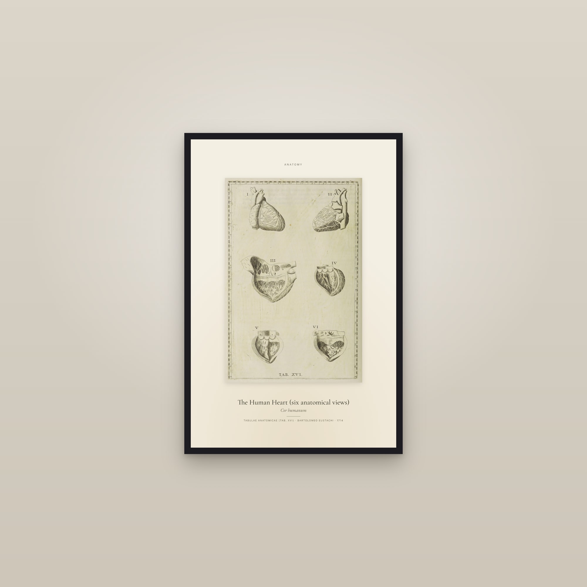
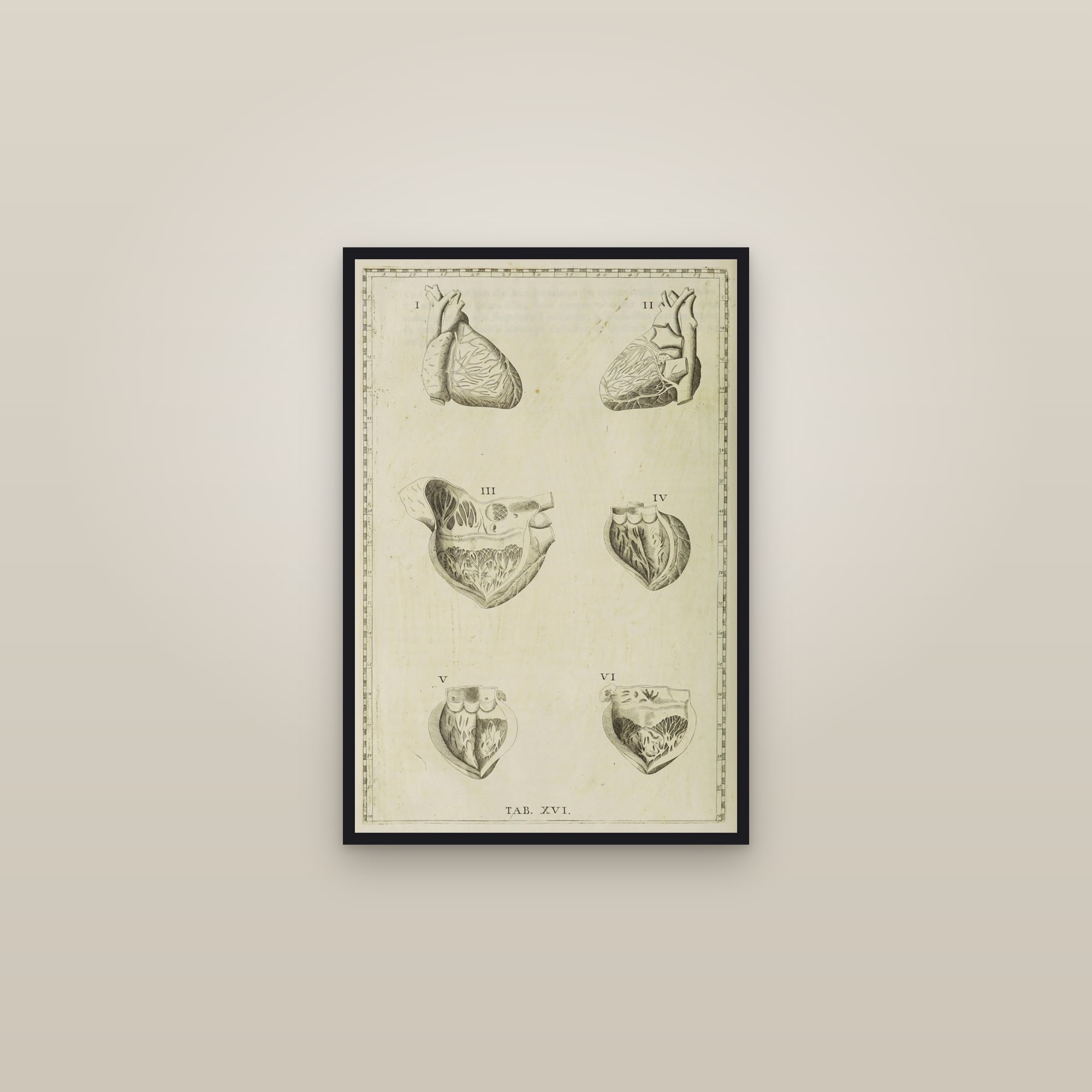
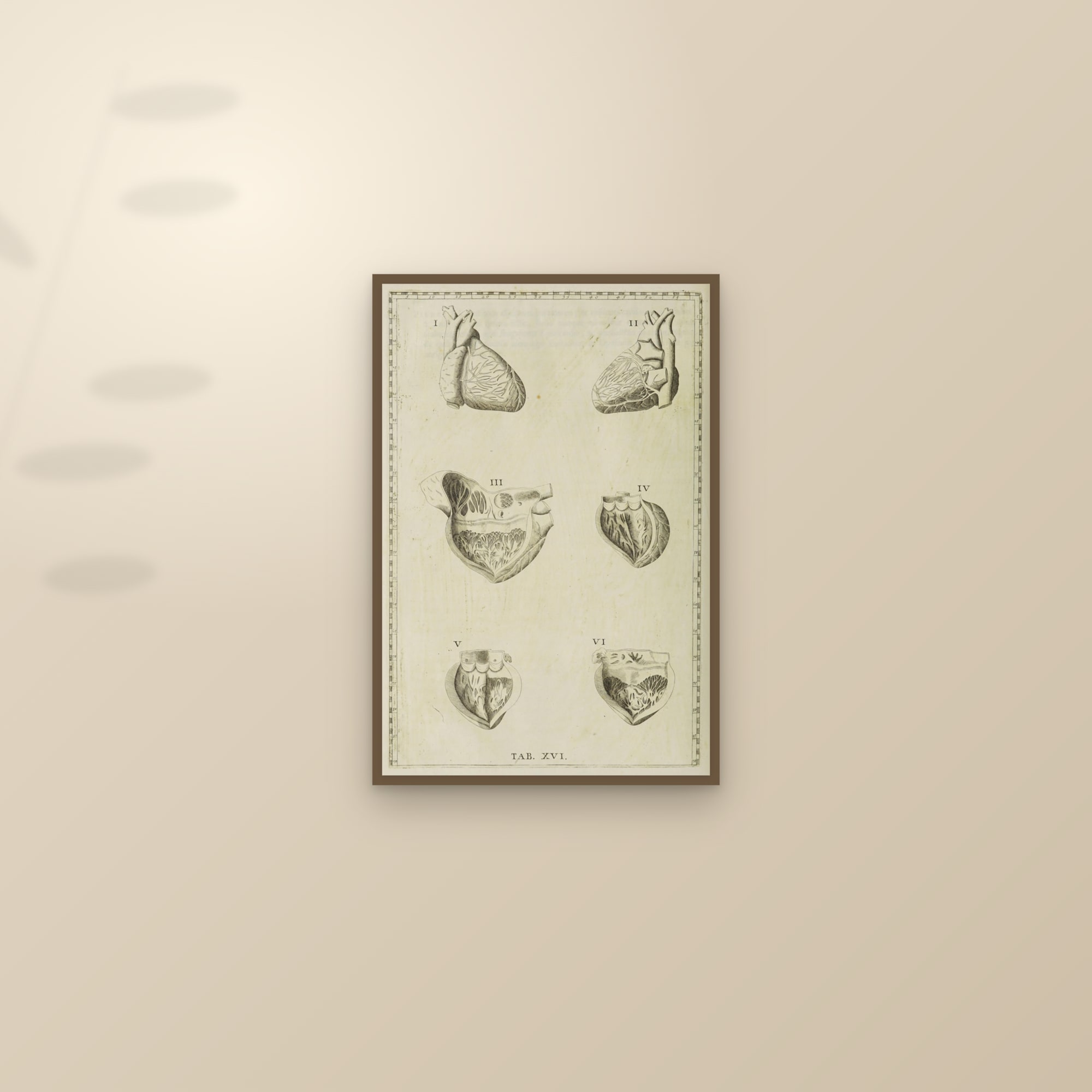
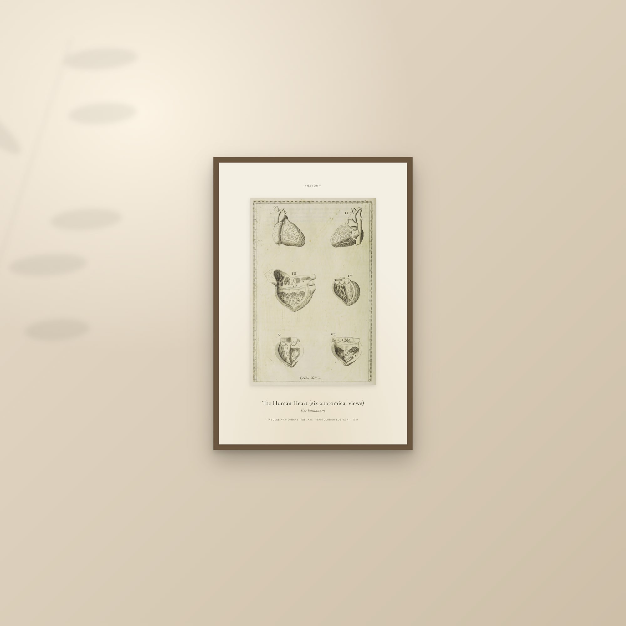
Restoring a plate means four things here, and nothing else: start from the best scan that exists, re-centre the plate inside even margins using its own paper, crop what the scanner added, and rotate what the atlas printed sideways. It does not mean repainting a line, inventing a colour, cleaning the foxing off the sheet, or running the file through upscaling software. The worked example is Tab. XVI of Bartolomeo Eustachi's Tabulae anatomicae, six views of the human heart, engraved on copper by 1552 and first printed in Rome in 1714. It was in the catalogue twice, from two different scans, and the second time we did it properly.
 Before
 After
Step 1: start from the right scan
A library scan of a 1714 book comes in two kinds. A bitonal scan stores one bit per pixel, black or white, nothing between; libraries made millions for text pages, where they are fine. A continuous-tone scan stores the greys. On an engraving the difference is the whole image. The engraver of this plate, Giulio de' Musi according to a 1742 preface cited in the Wellcome Collection's record, modelled the heart with cross-hatching: fine parallel cuts whose spacing carries the shading. From a metre away the hatching is a grey. A bitonal threshold must call each pixel black or white, so close lines fuse into a black mass and shallow ones vanish. In the before image above, the opened ventricle of figure III is a black pool and the walls of figures I and II go flat; the paper is pure white; the numbered ruler is a jagged band.
Most copies of this plate online come from a scan like that. Ours comes from the Wellcome Collection's scan of its copy of the 1714 Tabulae anatomicae (Rome, Francesco Gonzaga, folio, 47 plates), published under a Public Domain Mark, which the record glosses as "You can use this work for any purpose without restriction under copyright law." The same scan sits on the Internet Archive under the Wellcome Library's identifier. According to the Wellcome record of the plates' history, Eustachi had 46 plates ready by 1552, died in 1574 without publishing most of them, and the papal physician Giovanni Maria Lancisi found the coppers with the heirs of Eustachi's collaborator Pier Matteo Pini and printed them in 1714 with a text of his own. The National Library of Medicine's note on Eustachi explains the ruler: the plates carry printed, numbered rulers on their edges so that the text can point to any structure by its coordinates. On a bitonal scan those numerals are barely legible; on the Wellcome scan they read 5 to 55 across the top and 5 to 90 down the side.
Step 2: look at every plate at 100 %
Nothing about a raw library scan is checked by software alone. Every plate is opened at full size and measured, because a bound volume is never scanned square: on the Wellcome scans the heart plate's frame sits 13.5 % to one side of the sheet, the arteries with arms raised sit 13.6 % high, most bordered plates are off by 3 % or more, and on some the ruler touches the cut edge. A contact sheet of thirty thumbnails shows none of this. A thumbnail is not evidence.
The same full-size pass reads each plate against its own engraving: that the title matches what the figure shows (a "front" view that is in fact a back view is a common cataloguing slip), that a page the atlas printed sideways is turned upright, that a library footer is cut, that an edge-to-edge crop never takes a figure's head. One person, every plate, at 100 %.
Step 3: find the frame, with a threshold set per plate
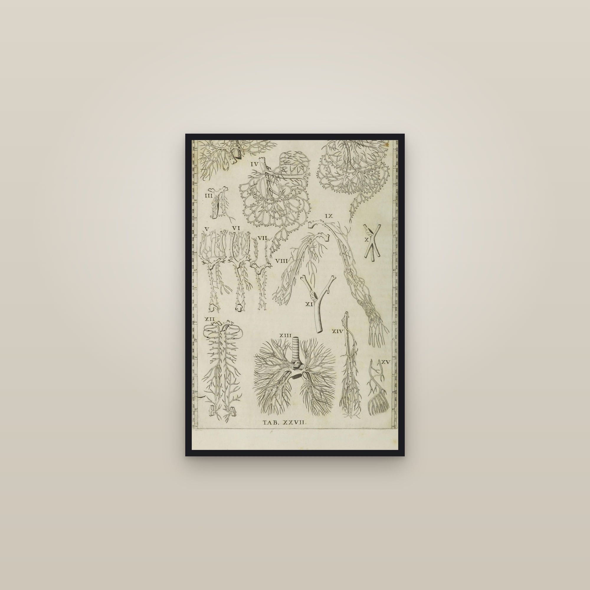
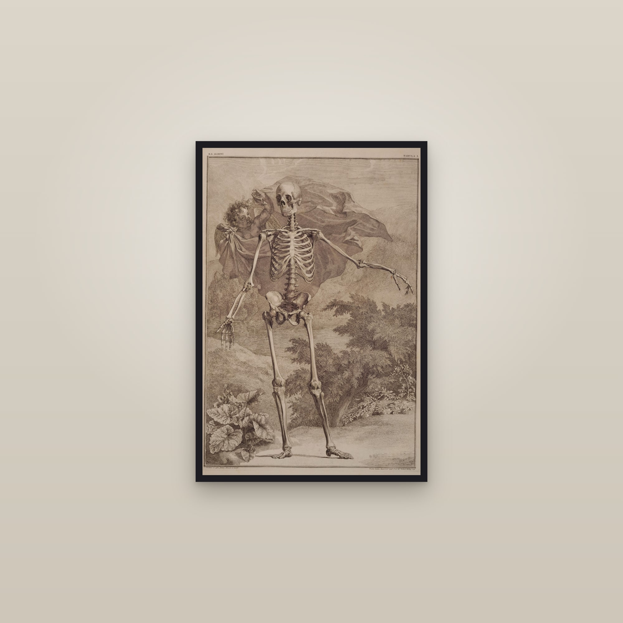
A Eustachi or Albinus plate has a ruled frame, and the restoration begins by finding it exactly. The script reads the plate at native resolution and looks for ink: a pixel is ink when its luminance is more than 45 units below the paper's. The threshold has to be per plate. The Wellcome paper carries show-through of the text on the other side of the leaf (visible at the top of the heart plate, between figures I and II), and a fixed threshold read that ghost text as frame. The rules themselves are a faint grey, luminance 150 to 180 on a 0 to 255 scale, not black. Measure the ink profile first, then choose the threshold.
Step 4: re-centre to an even 2.5 % margin, from the plate's own paper
With the frame's edges known, the target is a margin of 2.5 % of the frame's width outside it, on all four sides. For each side there are three cases.
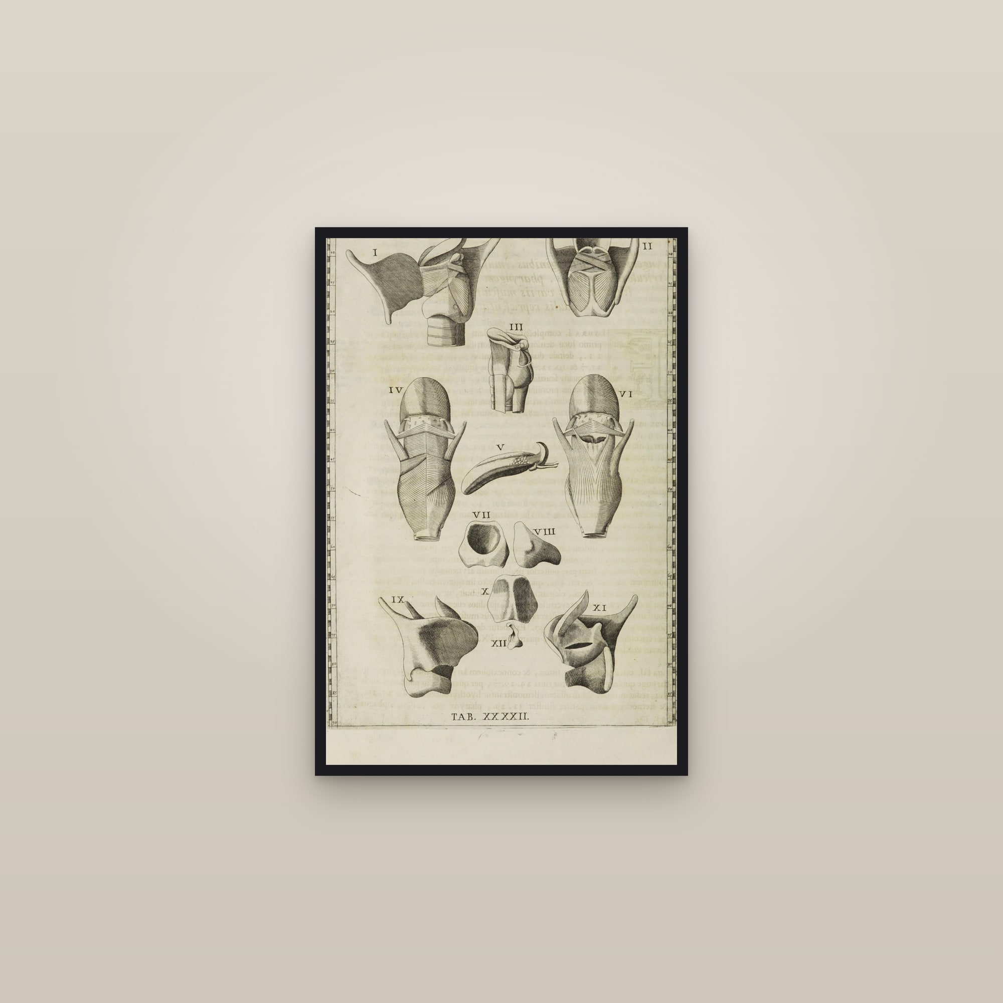
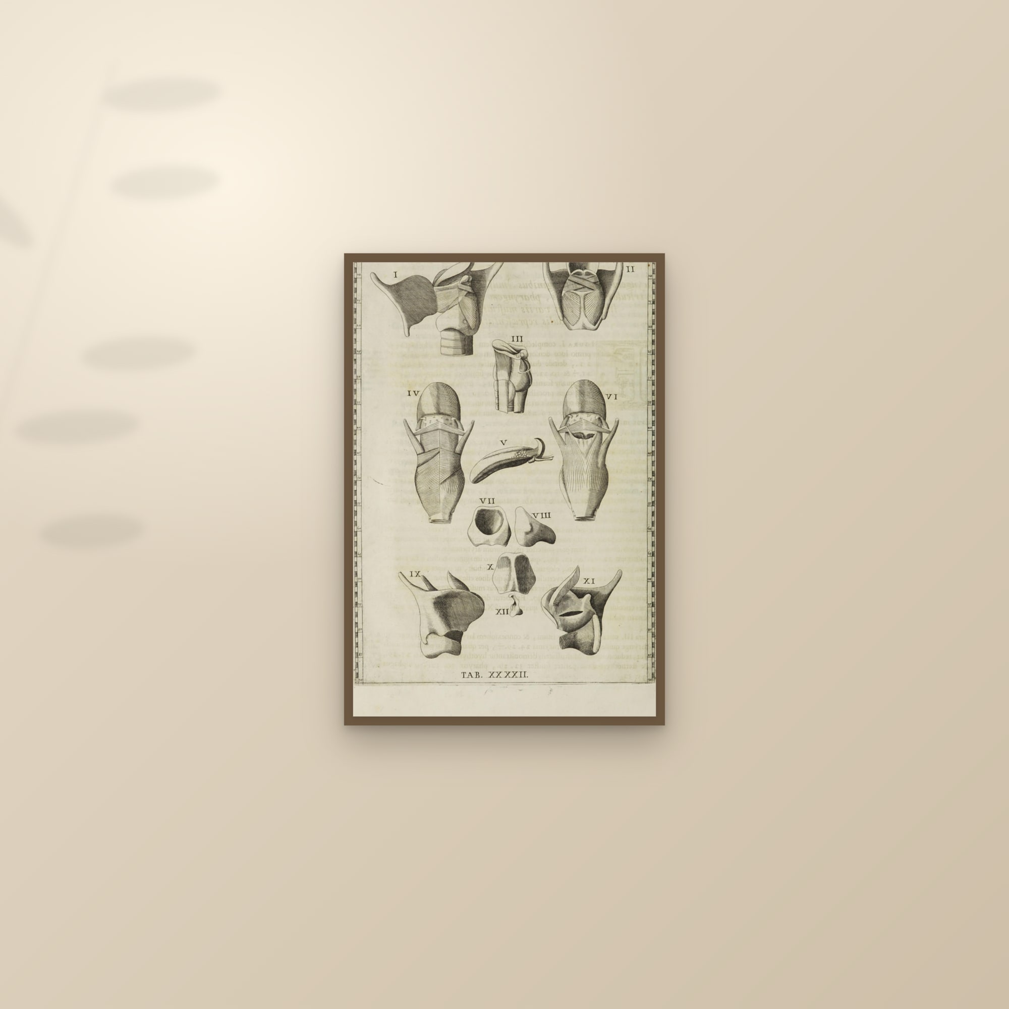
Where the scan has at least 12 pixels of real paper beyond the frame, the page is cropped or mirror-padded to the target: more paper than 2.5 % is cropped; a little less is made up by mirroring a strip of the plate's own paper from that edge. Where the scan clipped the frame itself, fewer than 12 pixels of paper, both sides of that axis are set flush, so the plate is at least symmetrical. The larynx plate below is one of two cut that way: the paper outside the left and right rulers was cut on both sides rather than one.
What the script never does is paint paper. Synthetic paper, whether a flat fill, a texture or sampled noise, reads at 100 % as a rectangle of slightly wrong cream next to real paper with real fibres and real foxing. The margin is always the page's own. Fifty-seven plates (39 Eustachi, 18 Albinus) went through this and are centred within half a per cent.
Step 5: why the ruler band stays
For the edge-to-edge edition, the one that fills the sheet, a tighter crop to the inner rule of the frame would hide the ruler band. It is not done automatically, and the reason is worth knowing if you ever restore a plate yourself: on an engraving, a line detector cannot tell a tick mark, a numeral, a patch of hatching or the edge of a figure from the rule it is looking for, and whichever it picks, it cuts into the art. Thumbnails will look fine. The corners at 100 % will not.
So the edge-to-edge edition uses the re-centred plate as it is; the 2:3 sheet trims the ruler band symmetrically on the long axis, and the specimen edition shows the whole frame, ruler and all. If a plate ever needs a tighter cut, a person makes it by hand, looking at the corners.
Step 6: crop what the scanner added
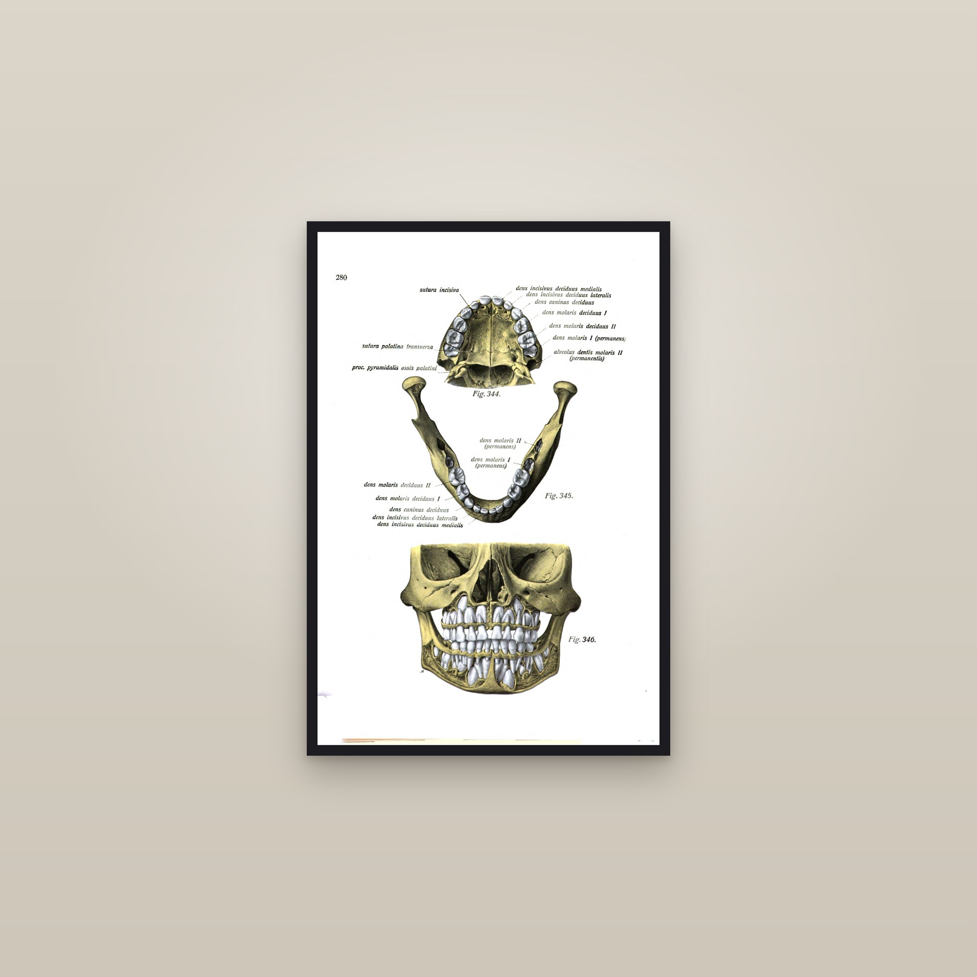
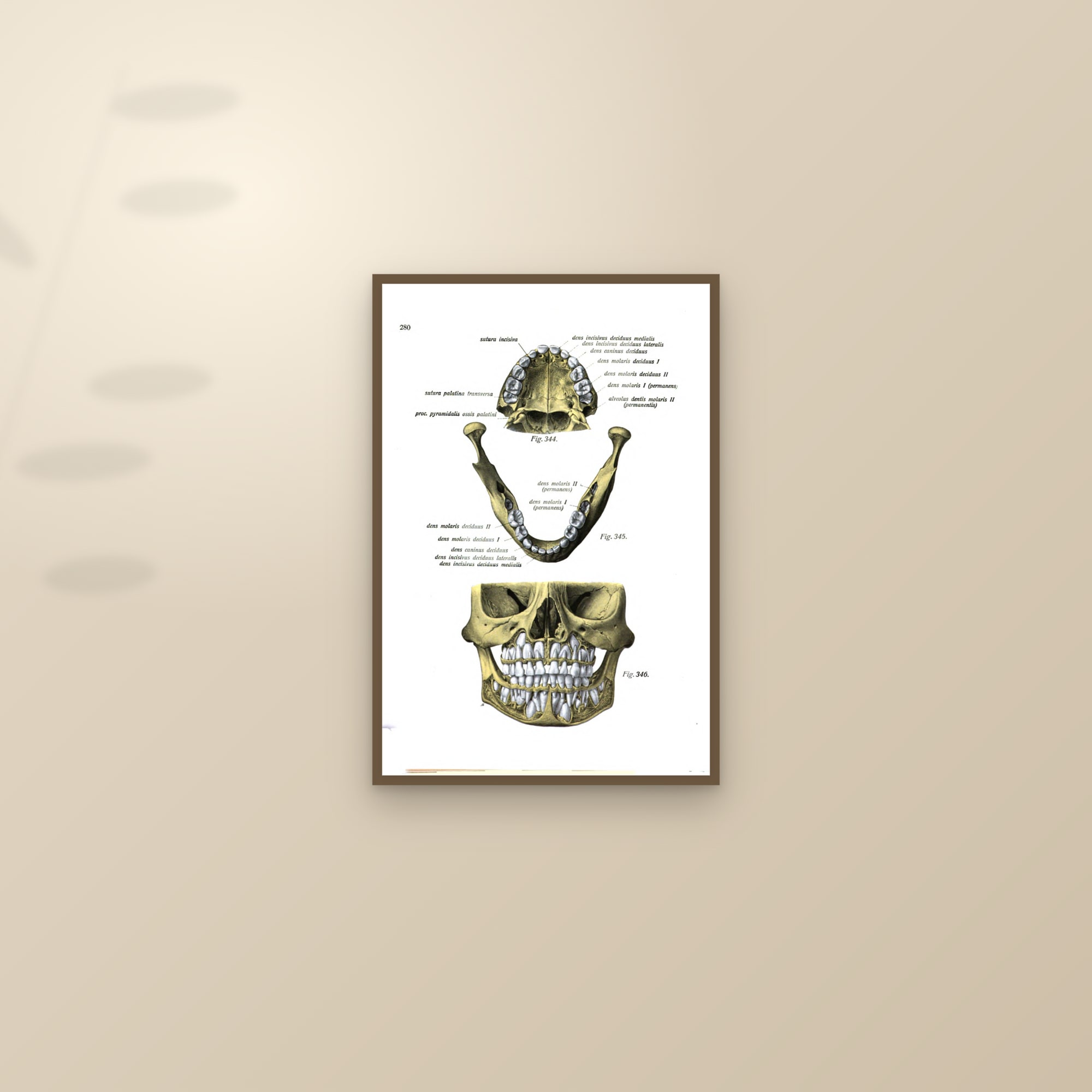
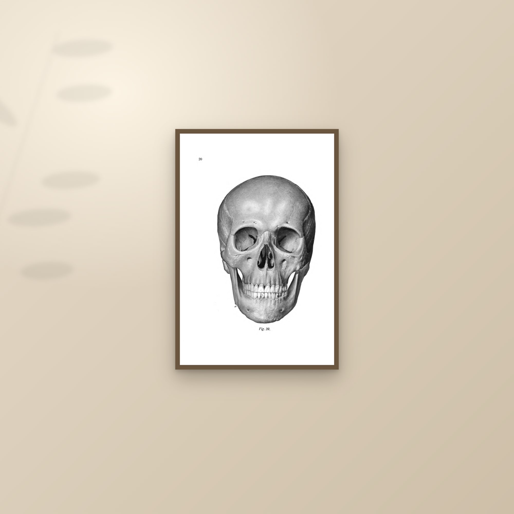
Scanners add things the printer did not. A page from Johannes Sobotta's Atlas der deskriptiven Anatomie des Menschen (1919), the skull from the front, carried a university library footer under the figure; the bottom 8 % of the page was cropped. The page of a child's jaws and teeth from the 1920 volume carried a yellow stripe down the left edge, 3.5 % of the width, and a scanner colour bar along the bottom, 4.5 % of the height; both cropped. The same category covers calibration strips on poster scans, the dark scanner board round them, and the grey studio wall in some museum photographs of a stretcher: detected, checked by eye, cut. The page number and the figure caption stay, because they were printed.

Step 7: rotate what the atlas printed sideways
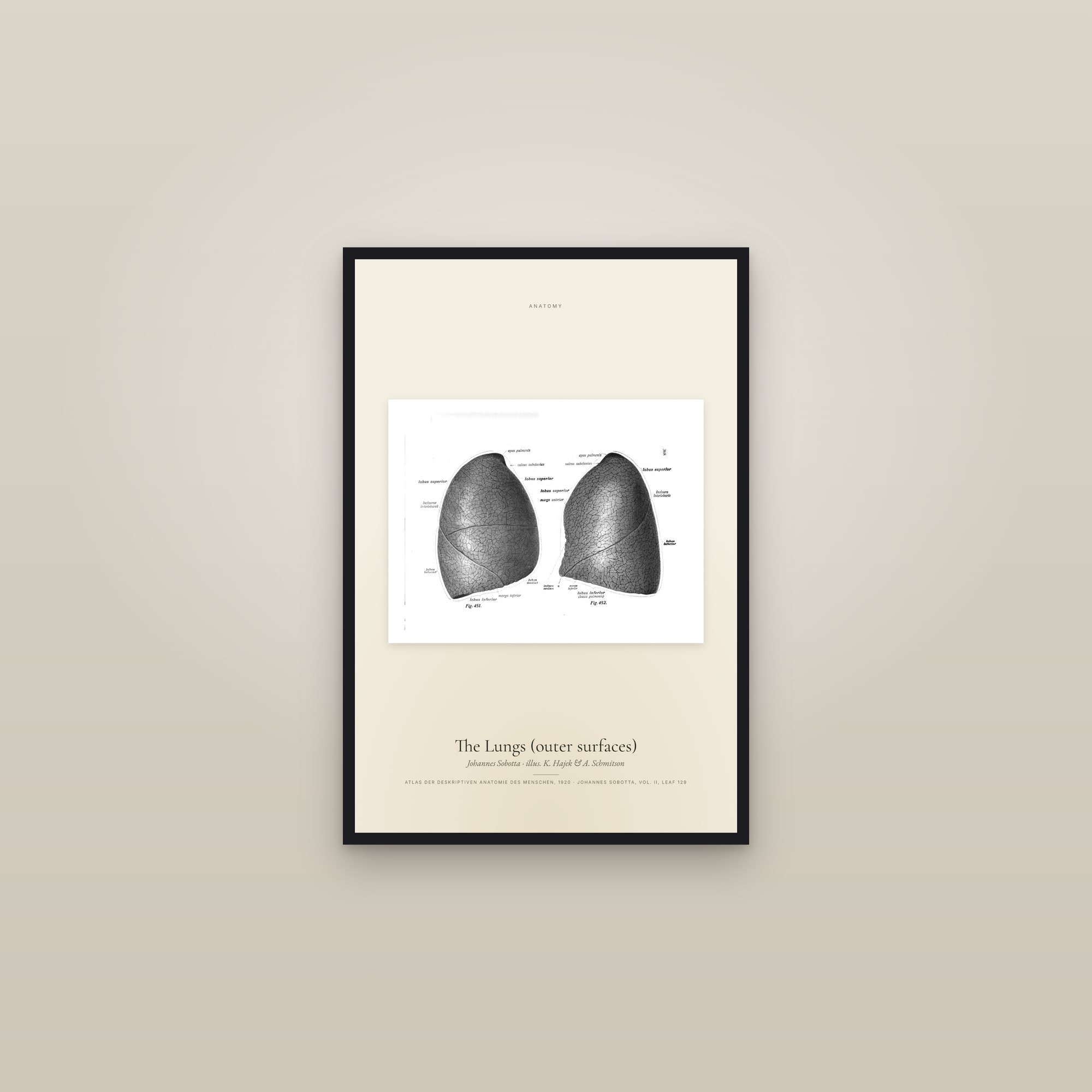
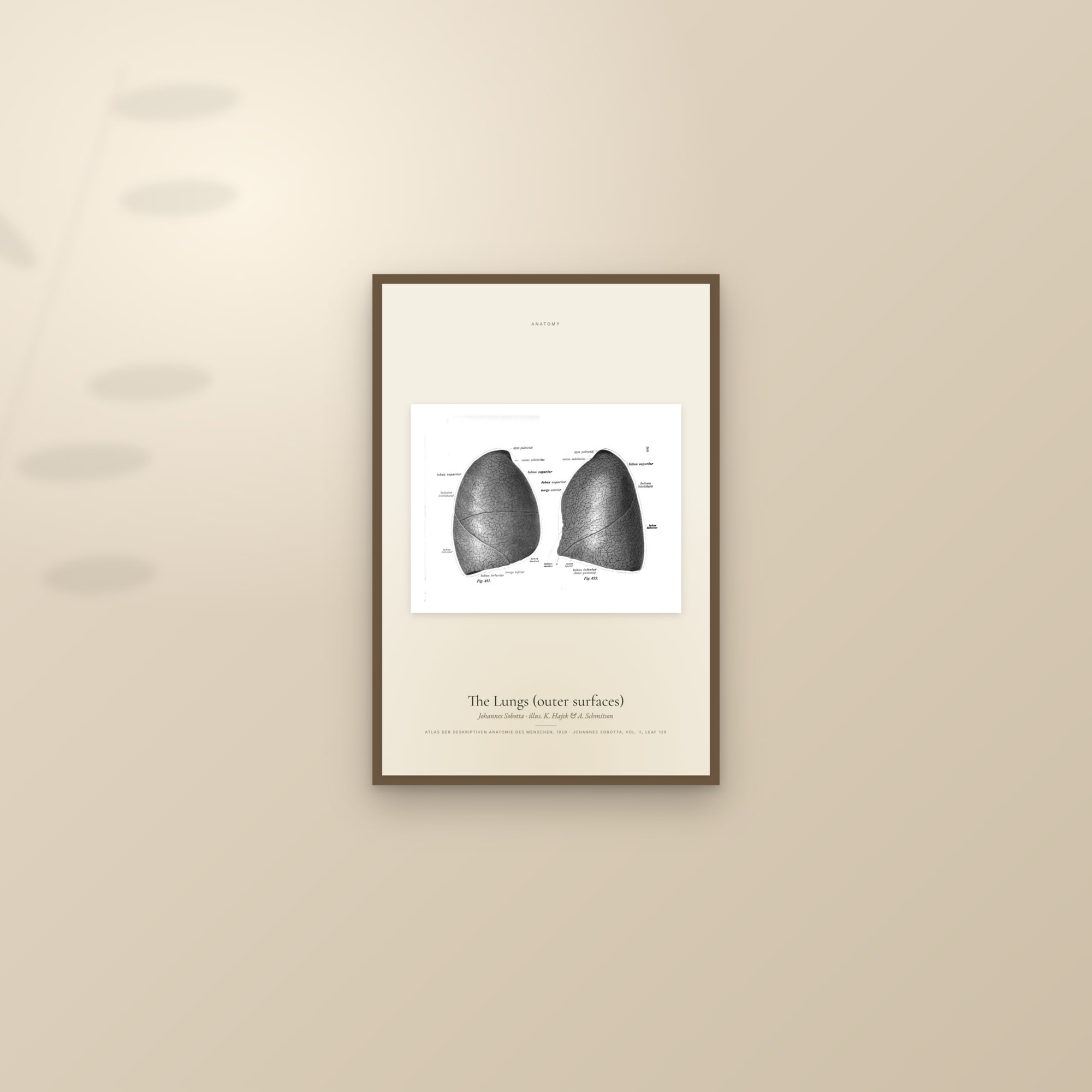
Sobotta set his wide figures across the page, so that the student turned the book. Seventeen of those pages, lungs, hand muscles, bones of the hand and foot, the heart opened and in section, the brain with its arteries, were rotated 90° to read upright; on every one the text had run from bottom to top, which is the tell. A rotated page is a landscape, never cover-cropped to the upright sheet because that slices the subject; each ships as a single specimen print, the whole page on a cream ground with a typeset label.
Step 8: head-safe trims for figures that sit high
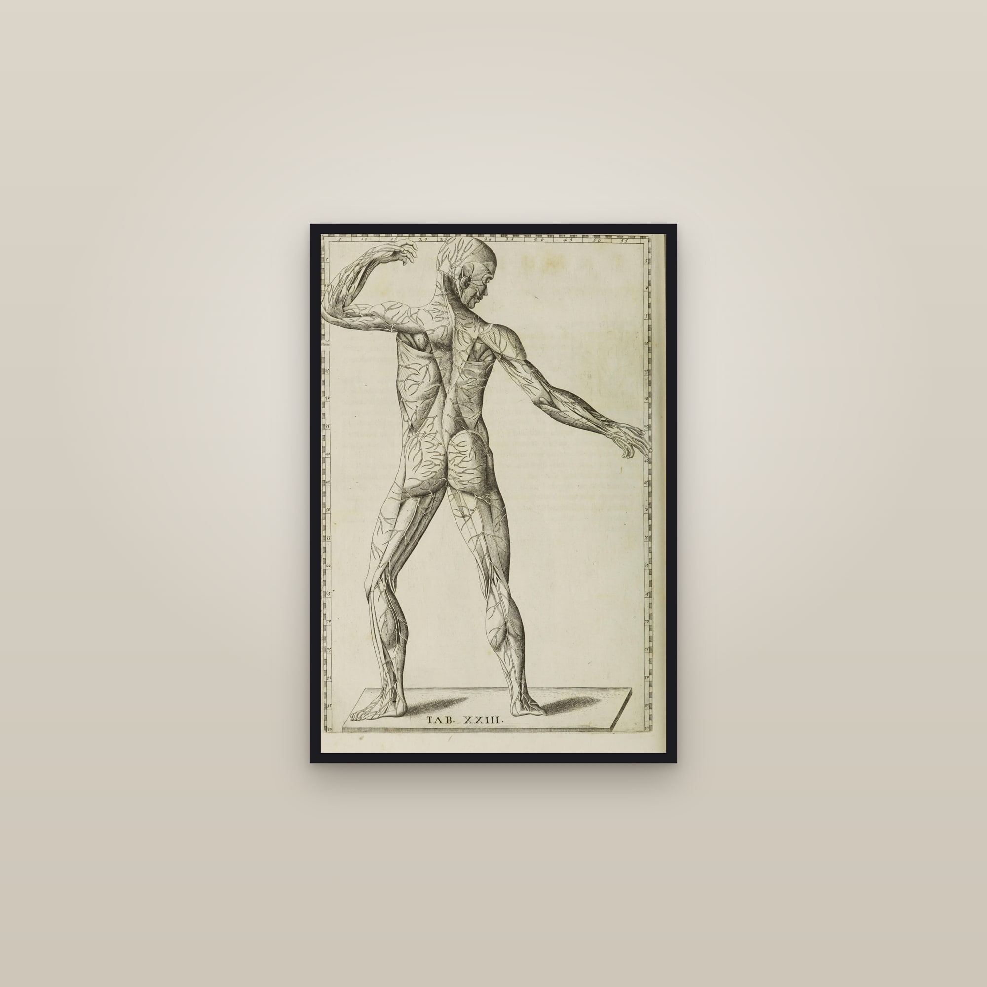
Three Eustachi figure plates, the arteries, the veins seen from the side, and a posterior muscle man, stand so tall on the page that a centred 2:3 crop would take the top of the head with the ruler. For those the edge-to-edge window is anchored at the top of the plate instead of the centre: it keeps the head and the top ruler and gives up the "TAB." caption line at the foot. The specimen edition keeps everything.
Step 9: check it again
After every pass the plates go onto a sheet at full size and the corners are zoomed to 100 %. Then the layout is rendered to a 7200 × 10800 pixel master, a mockup is made of the print framed on a wall, and that is looked at too, as a buyer would see it. The whole route from scan to sheet is in How a fine-art print is made.
What restoration is not
It is not repainting: no line in the heart plate was redrawn, thickened or cleaned. It is not colour: the cream is the cream Wellcome scanned; we do not tint, bleach or warm a sheet, and the foxing and the ghost of the verso stay, because they are the page. It is not sharpening or upscaling: a plate scanned at 2,656 pixels wide is printed from 2,656 pixels, which is why the heart plate stops at 16 × 24 inches rather than being sold at 24 × 36 with software guessing the missing detail (Print sizes and ratios explained has the arithmetic). What you buy is the 1714 impression, scanned by a library, centred and cropped by us, on 250 gsm archival paper, from $39.
Shop the art in this piece
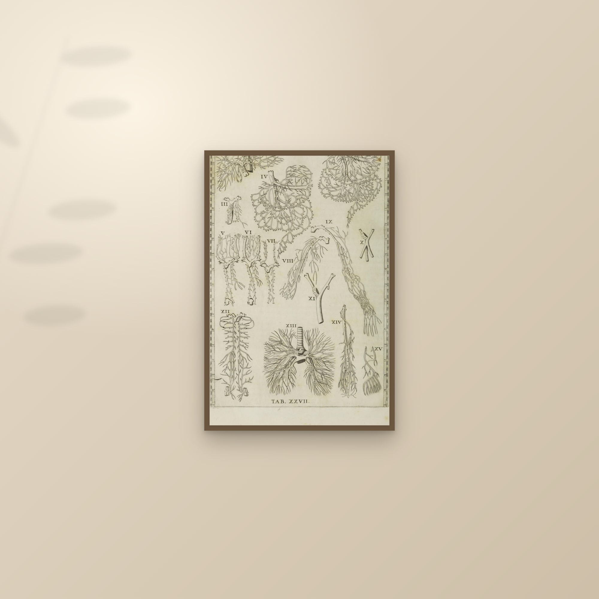
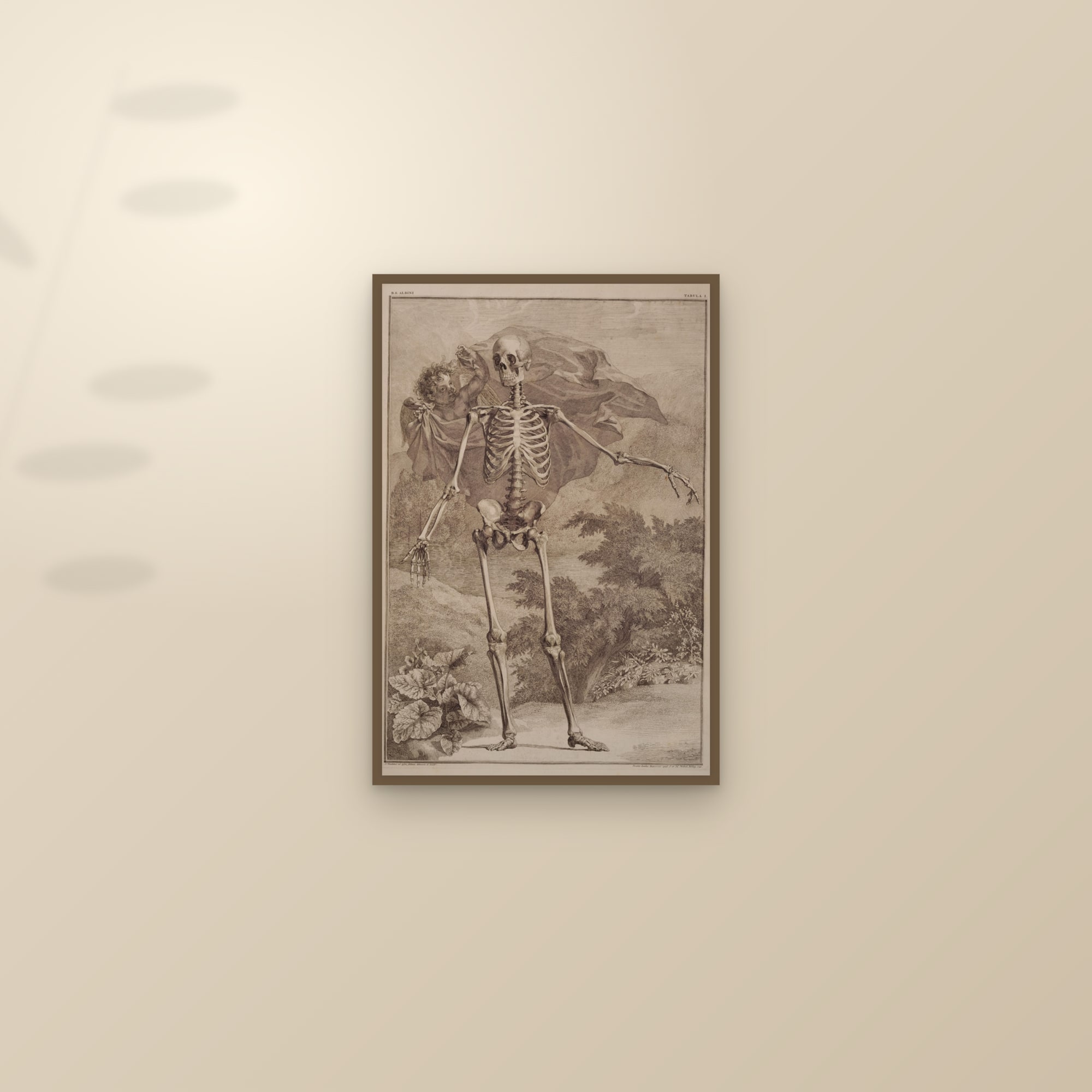
The same passes run on every scanned line: the fruit watercolours had their dark scanner backdrop shaved; the paintings have their corners checked for studio backdrop. For what the ink and paper are, see What is a giclée print?.
Questions people ask
What does Madten do to a plate before printing it?
Four things, and only these: pick a continuous-tone scan over a bitonal one; re-centre a bordered plate inside even margins using its own paper; crop scanner additions such as library footers and colour bars; rotate pages an atlas printed sideways. The artwork itself is not touched.
Do you repaint, recolour or sharpen old plates?
No. We do not repaint lines, invent colour, remove foxing or age-toning, or run upscaling software. What you see is the museum or library scan of the printed page, cropped and centred.
Why do two scans of the same Eustachi plate look so different?
Most copies online come from a bitonal scan, every pixel black or white, which merges the engraver's hatching and bleaches the paper. Ours comes from the Wellcome Collection's continuous-tone scan of the 1714 volume, with the frame re-centred on the sheet; on the raw scan it sits 13.5 % to one side.
What is a bitonal scan?
A scan saved with one bit per pixel: black or white, no greys. Libraries made them in bulk for text pages. On an engraving they destroy the shading, because cross-hatching is grey at a distance and a threshold either drops the lines or fuses them.
Where does the paper around a re-centred plate come from?
From the same scan. Where the page has real paper beyond the frame it is cropped to an even margin; where it has slightly less, a strip of the plate's own paper is mirrored to make up the difference. No synthetic paper is ever painted in; at 100 % it reads as a patch next to real fibres.
Sources
- Wellcome Collection: Tabulae anatomicae (Rome, 1714), 47 plates, Public Domain Mark
- Wellcome Collection: engraving from the Tabulae, with the history of the plates (de' Musi, Pini, Lancisi)
- Internet Archive: Wellcome Library scan of the 1714 Tabulae anatomicae (b3045279x), Public Domain Mark
- US National Library of Medicine, Historical Anatomies: Bartolomeo Eustachi
- Creative Commons: Public Domain Mark 1.0
Start hereHow a fine-art print is made: from a 1714 plate to your wall